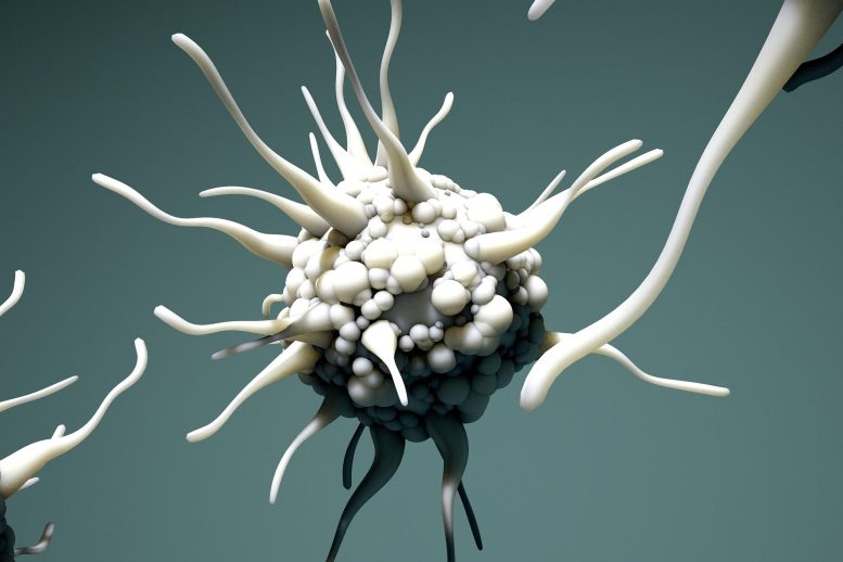
Researchers Have Created a New Assay To Detect Cellular Protein Secretion
Washington University researchers have developed the FluoroDOT assay, a revolutionary protein visualization technique that uses technology from the James Webb telescope. This technique measures proteins secreted by a single cell in 30 minutes using plasmonic-fluor, an ultrabright nanoparticle. The assay allows detailed visualization of cell and protein secretions and has been validated with proteins secreted from human and mouse cells.
The James Webb telescope has just been able to capture breathtaking photos of faraway galaxies that were previously only seen as hazy patches. The James Webb version for observing single-cell protein secretion has been created by Washington University in St. Louis researchers using a revolutionary technique that allows for stunningly detailed visualization of the proteins produced by cells.
The researchers, led by Srikanth Singamaneni, the Lilyan & E. Lisle Hughes Professor of Mechanical Engineering & Materials Science at the McKelvey School of Engineering, and Anushree Seth, a former postdoctoral fellow in Singamaneni’s lab, developed the FluoroDOT assay. The study was recently published in Cell Reports Methods. The highly sensitive assay is able to see and measure proteins secreted by a single cell in about 30 minutes.
Together with scientists from other universities and the Washington University School of Medicine, they discovered that the FluoroDOT assay is versatile, affordable, adaptable to any laboratory setting, and has the potential to provide a more comprehensive picture of these proteins than the currently commonly used assays. Biomedical researchers turn to secreted proteins for information on cell-to-cell communication, cell signaling, activation, and inflammation, among other actions, but present approaches are restricted in sensitivity and may take up to 24 hours to process.
The Role of Plasmonic-Fluors in Enhancing Fluorescence
The plasmonic-fluor, a plasmon-enhanced nano label created in Singamaneni’s lab that is 16,000 times brighter than conventional fluorescence labels and has a signal-to-noise ratio that is over 30 times greater, is what distinguishes the FluoroDOT assay from other assays.
“Plasmonic-fluors are composed of metal nanoparticles that serve as antennae to pull in the light and enhance the fluorescence emission of molecular fluorophores, thus making it an ultrabright nanoparticle,” Singamaneni said.
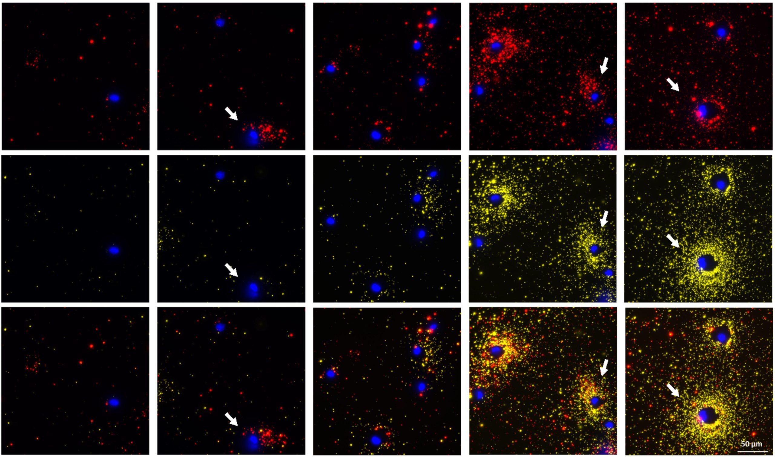
This ultrabright emission of plasmonic-fluor allows the user to see extremely small quantities of secreted protein, which they are unable to do in existing assays, and measure the high-resolution signals digitally using the number of particles, or dot pattern, per cluster, or spot, using a custom-built algorithm. In addition, it doesn’t require special equipment. Singamaneni and his collaborators first published their work with the plasmonic-fluor in Nature Biomedical Engineering in 2020.
The patent-pending plasmonic fluor technology is licensed by the Office of Technology Management at Washington University in St. Louis to Auragent Bioscience LLC.
“Using a simple fluorescence microscope, we are able to simultaneously image a cell along with the spatial distribution of the proteins secreted around it,” said Seth, who worked on this project as a postdoctoral scholar in Singamaneni’s lab and continues to work on it as a principal scientist (cellular applications) for Auragent Bioscience. “We saw interesting secretion patterns for different cell types. This assay also enables concurrent visualization of two types of proteins from individual cells. When the multiple cells are subjected to the same stimuli, we can distinguish the cells that are secreting two proteins at the same time from the ones that are only secreting one protein or are not secreting at all.”
Real-World Applications and Validation
To validate the technology, the team used proteins secreted from both human and mouse cells, including immune cells infected with Mycobacterium tuberculosis.
One of the collaborators and co-authors, Jennifer A. Philips, MD, Ph.D., the Theodore and Bertha Bryan Professor in the departments of Medicine and Molecular Microbiology and co-director of the Division of Infectious Diseases in the School of Medicine, has used the FluoroDOT assay in her lab.
“When Mycobacterium tuberculosis infects immune cells, those cells respond by secreting important immune proteins, called cytokines,” Philips said. “But not all cells respond to infection the same way. The FluoroDOT assay allowed us to see how individual cells in a population respond to infection — to see which cells are secreting and in which direction. This was not possible with the older technology.”
Reference: “High-resolution imaging of protein secretion at the single-cell level using plasmon-enhanced FluoroDOT assay” by Anushree Seth, Ekansh Mittal, Jingyi Luan, Samhitha Kolla, Monty B. Mazer, Hemant Joshi, Rohit Gupta, Priya Rathi, Zheyu Wang, Jeremiah J. Morrissey, Joel D. Ernst, Cynthia Portal-Celhay, Sharon Celeste Morley, Jennifer A. Philips and Srikanth Singamaneni, 5 August 2022, Cell Reports Methods.
DOI: 10.1016/j.crmeth.2022.100267
The study was funded by the National Institutes of Health.
Never miss a breakthrough: Join the SciTechDaily newsletter.
Follow us on Google and Google News.