
MRI scans often have difficulty producing clear images of deep or delicate tissues. Researchers at the Max Delbrück Center report that a new lightweight antenna can improve image quality and may reduce scan times without requiring changes to existing MRI machines.
Magnetic resonance imaging (MRI) is among the most important tools doctors use to diagnose disease. Yet some tissues located deep in the body, including brain regions and delicate eye and orbit structures that are especially important in ophthalmology, remain difficult to capture in sharp detail. The limitation comes not from the MRI scanner itself, but from the hardware responsible for transmitting and receiving radio signals.
A team led by Nandita Saha, a doctoral student in the Experimental Ultrahigh Field Magnetic Resonance lab of Professor Thoralf Niendorf at the Max Delbrück Center, has now created an MRI antenna based on advanced materials that addresses this problem. The device can produce clearer images in less time and can be used with MRI machines already in place. The work was published in Advanced Materials.
Niendorf and his colleagues collaborated closely with researchers at Rostock University Medical Center, bringing together MRI physics, clinical ophthalmology, and translational imaging. The Rostock team is also helping evaluate the technology for clinical use.
“By using concepts from metamaterials, we were able to guide radiofrequency fields more efficiently and demonstrate how advanced physics can directly improve medical imaging,” says Niendorf, senior author of the paper. “This work shows a pathway toward faster, clearer MRI scans that could benefit patients in many clinical areas.”
Rethinking MRI hardware with metamaterials
MRI creates images by sending radiofrequency (RF) signals into the body and measuring how tissues respond within a powerful magnetic field. Stronger signals produce better images. Standard MRI antennas, also known as RF coils, often have trouble gathering enough signal from deep tissues or regions with complex anatomy. As a result, scans can take longer, and images may miss fine details.
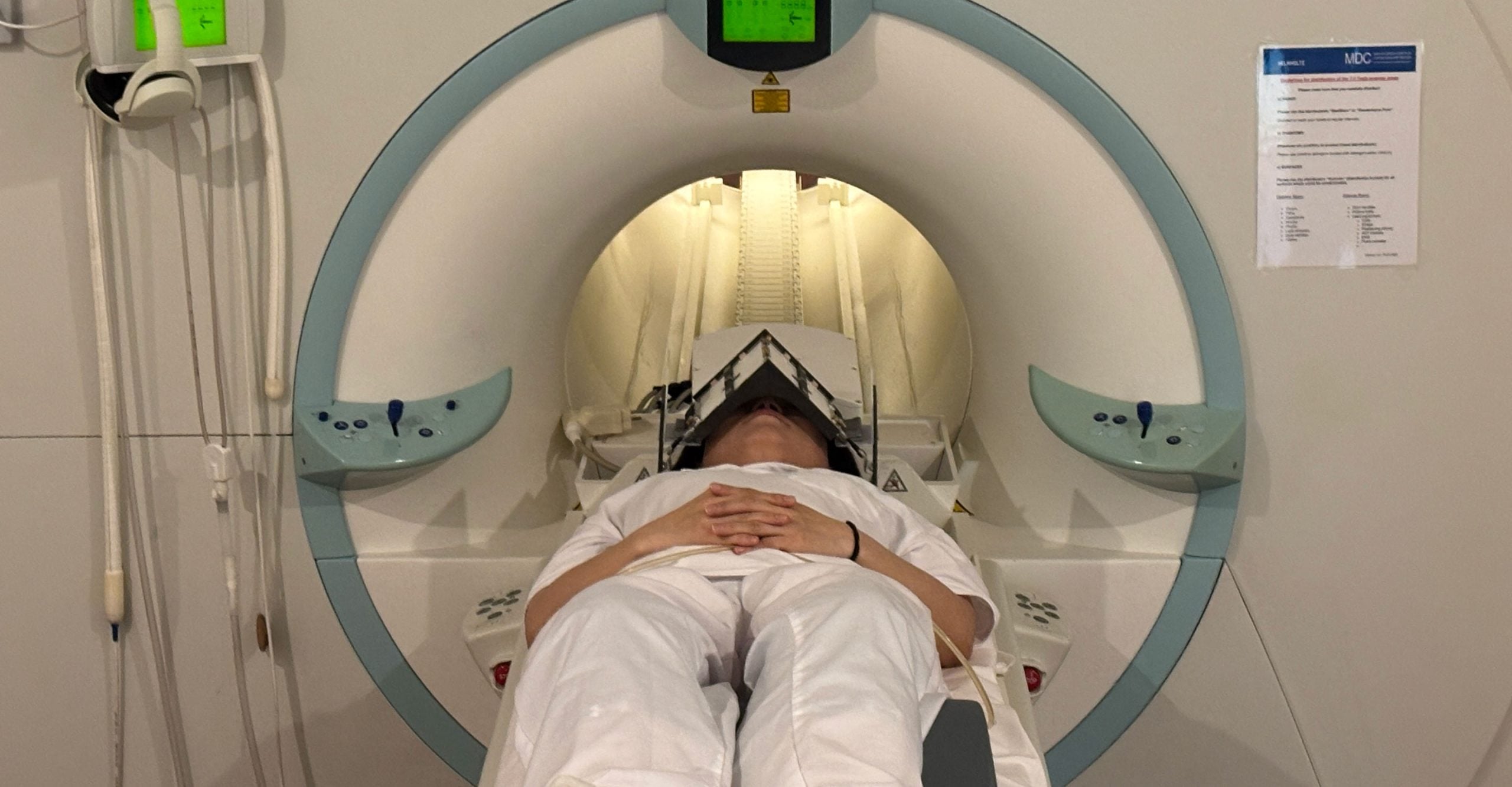
To solve that problem, the researchers built metamaterials directly into the MRI antenna. Metamaterials are designed structures that manipulate electromagnetic waves in ways natural materials cannot. The engineered RF antenna strengthens signals from targeted tissues, increases spatial resolution, improves image clarity, and speeds up data collection. Importantly, it works with existing MRI systems, so hospitals would not need entirely new scanner infrastructure. The team tested the technology by imaging the eye and orbit region in volunteers at 7.0 Tesla.
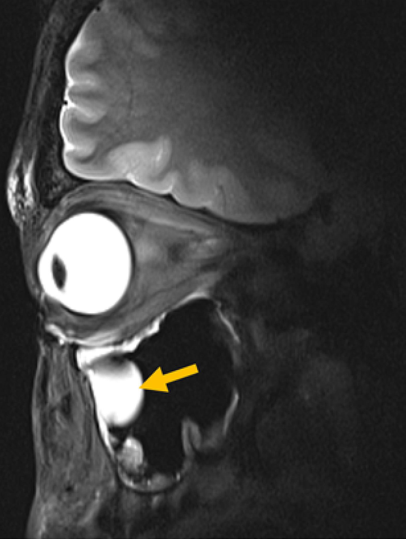
“Our research demonstrates clear relevance for ophthalmological applications as it can facilitate anatomically detailed, high-spatial resolution MRI of the eye,” says Professor Oliver Stachs, a co-author of the paper at University Medicine Rostock. “It offers the potential to open a window into the eye and into (patho)physiological processes that in the past have been largely inaccessible.”
“Our goal was to rethink MRI hardware from the modern physics of antenna design,” adds Saha. The same technology could also be adjusted to protect sensitive body regions during MRI, such as reducing unwanted heating around medical implants, she adds. It may also help concentrate RF energy more precisely for MRI-guided therapies used in cancer care, including gentle heating of tumors (hyperthermia) or thermal ablation of tissue.
Better diagnostics
MRI exams can be uncomfortable and lengthy for patients, especially when scans must be repeated because important structures are not visible enough. Shorter scans would reduce the time patients spend inside the machine. Sharper images could help physicians make diagnoses with greater confidence. Because the new antenna is compact and lightweight, it can also be shaped for specific body regions, potentially making scans more comfortable.
Niendorf says the technology could be adapted for MRI systems operating at magnetic field strengths below or above 7.0 T, for imaging anatomy beyond the eye, orbit, or brain, and for tracking metabolism or drug movement inside the body. Specialized MRI methods that detect atoms such as sodium or fluorine could also use the technology to capture stronger signals and clearer images, he adds.
“Innovations in imaging hardware have the potential to transform diagnostics, and this study is an important step toward next-generation MRI technology,” says Dr. Ebba Beller, a co-author of the paper at Rostock University Medical Center.
The researchers are now preparing larger studies at several hospitals and modifying the design for other organs, including the heart and kidneys. The collaboration will continue through long-standing reciprocal visiting scientist appointments held by Stachs and Niendorf.
Reference: “Metamaterial Antennas Enhance MRI of the Eye and Occipital Brain” by Nandita Saha, Bilguun Nurzed, Mostafa Berangi, Andre Kuehne, Helmar Waiczies, Igor Fabian Tellez Ceja, Xiang Hu, Thomas Gladytz, Lisa Krenz, Dave Huebler, Beate Endemann, Claudia Brockmann, Ebba Beller, Oliver Stachs and Thoralf Niendorf, 2 February 2026, Advanced Materials.
DOI: 10.1002/adma.202517760
This project was funded by the DFG project as a joint collaboration between the Max Delbrück Center and the Medical University Rostock.
Never miss a breakthrough: Join the SciTechDaily newsletter.
Follow us on Google and Google News.
3 Comments
thanks for this
thanks
Is this a T mri used in level 4 epilepsy centers or different tech can someone explain differences? Ty