A new virus killing superworms was discovered by Rutgers scientists using cryo-electron microscopy, offering insights into pathogen identification and potential outbreak management.
Scientists from Rutgers University-New Brunswick have discovered a virus that has caused a nationwide die-off of superworms, which are a dietary staple for many pets and increasingly used as an alternative protein source for humans. With this discovery, reported in a recent Cell article, they have devised a method to search for and identify emerging viruses and pathogens in plants, animals, and humans.
Innovative Research Techniques in Virology
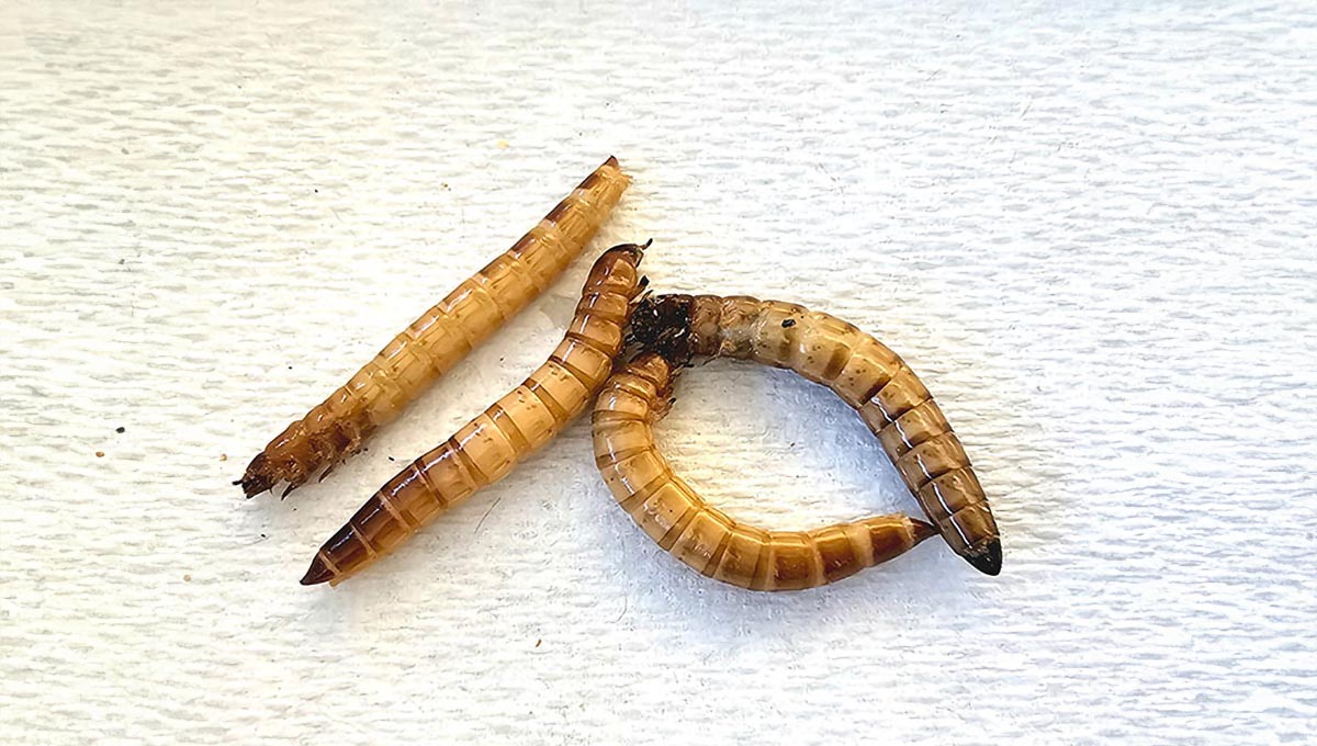
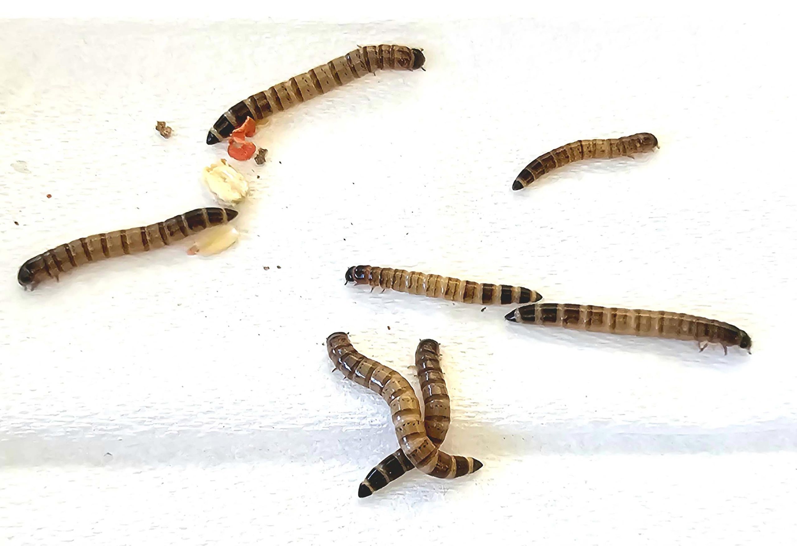
Using chopped-up beetle carcasses forming a slurry and an electron microscope cooled by liquid nitrogen, the scientists discovered what they have titled Zophobas morio black wasting virus. The name is derived from the virus’ deadly effect on a species of darkling beetle, Zophobas morio, native to the subtropics, particularly in the insect’s immature larval stage when it emerges from its eggs as large, brown superworms. This species was named “superworm” because its larvae are bigger, at about 2 inches in length, than any others grown as feed.

Unraveling the Superworm Mystery
Jason Kaelber, an author of the study and an associate research professor at the Institute for Quantitative Biomedicine (IQB) at Rutgers-New Brunswick, worked with Judit Penzes, the first author of the study and a postdoctoral associate at IQB.
“Judit was looking to identify the reason beetle farmers were losing all their superworm colonies to a deadly disease and I was looking to develop ways of discovering new viruses that don’t depend on DNA or RNA sequencing,” Kaelber said. “We ended up discovering the virus that has been sweeping the country and killing superworms.”
The scientific investigation began more than a year ago, when Penzes, a molecular virologist, was contacted by beetle farm owners whose superworms were mysteriously dying off at alarming rates. Penzes was already well known in the industry because of earlier work where she isolated a virus that was killing crickets, another popular food for pets.
Investigative Techniques in the Lab
She started by collecting superworms at pet stores in New Jersey. “Whenever I went to a pet store, I immediately went to the feeder insect section, opened the containers and looked at the worms,” she said. “They were all infected. I told the owners of the stores what I was seeing that I was researching this virus, and asked if I could have the container. They were immediately on board. They told me to take as many as I needed.”
She returned to her lab, took a Magic Bullet blender, dropped the worm carcasses in, and blended them at a high speed. The process created a slurry of beetle juice which she took and processed using a virus purification method that separates the virus out due to its density. In the final step, she shined a fluorescent light on the centrifuge tube and the virus glowed blue.
“I said, ‘I got you,’ when I saw it,” Penzes said. “I knew then it was, indeed, a virus.”
Cryo-Electron Microscopy and Pathogen Identification
Next, Penzes worked with Kaelber, a fellow electron microscopist, to examine the virus using a cryo-electron microscope, which allows a three-dimensional view of the virus, including its interior.
“You’re taking a virus, a protein, a cell, etc., and you’re freezing it so quickly that the water solidifies without turning into ice crystals,” Kaelber said. “We actually can figure out what the amino acid sequence of the protein is without analyzing the DNA, and just by looking at that 3D structure, because we have such sharp resolution.”
They compared the structure of the protein with all known proteins using the database of the Protein Data Bank hosted at Rutgers and found that it is similar to a virus affecting cockroaches, but not identical, and part of a family of animal viruses known as parvoviruses.
“It’s a new one, different from anything that’s been sequenced or imaged before,” Penzes said.
Future Applications and Implications
The scientists are also grateful to superworm farmers nationwide who sent samples voluntarily, once word of the study got out. “The eagerness of the farmers to help us out researching the virus had an enormous role in helping this published study to be born,” Penzes said.
The effort, Kaelber said, provided a “proof of concept” that cryo-electron microscopy can be employed to directly discover and characterize new pathogens.
“In the future, if there’s ever a really important outbreak, we’re going to want to throw every tool we can at it to see what we can find,” Kaelber said. “We’d like to make diagnostic cryo-electron microscopy routinized, so that when there’s some unknown infectious disease, we have a lot of options for same-day identification of the causative agent.”
Advancements in Diagnostic Techniques
Cryo-electron microscopy has gained popularity in recent years, becoming a more prevalent method for 3D analysis of known specimens. However, the Rutgers work represents the first time the method was used on an unknown pathogen.
Concluding Remarks on Virus Research Impact
After discovering the virus, the researchers tested a way to protect the Z. morio beetles from disease, by injecting a closely related virus from another species that doesn’t cause symptoms. They are developing a vaccine based on that work.
“The discovery is important for two reasons,” Kaelber said. “First, beetle farmers can use this information to protect their colonies and understand which actions will be effective or ineffective at managing the epidemic. Second, the beetle epidemic was a real-world test of the technology that we hope can be useful to rapidly investigate future outbreaks in humans, plants or animals.”
Scientists Martin Holm of the Rutgers Institute for Quantitative Biomedicine and Samantha Yost of REGENXBIO Inc., in Rockville, Md., also authored the study.
Reference: “Cryo-EM-based discovery of a pathogenic parvovirus causing epidemic mortality by black wasting disease in farmed beetles” by Judit J. Penzes, Martin Holm, Samantha A. Yost and Jason T. Kaelber, 28 August 2024, Cell.
DOI: 10.1016/j.cell.2024.07.053
Never miss a breakthrough: Join the SciTechDaily newsletter.
Follow us on Google and Google News.
2 Comments
“she shined a fluorescent light on the centrifuge tube and the virus glowed blue.”
Surely that should be “shone”.
Languages evolve. English is regularizing its irregular past-tense verbs, like dove and sang or sung, to dived and even singed The article should still be in something like AP style for publication, and both “shined” and “glowed” would be considered awkward. As a scientific article, the correct terms would be “Illuminated” and “fluoresced”. Of course, “shined a fluorescent light” probably meant an ultraviolet light, as a regular fluorescent tube would just show the color it actually was, so it’s probably just poorly written by someone who also didn’t understand what happened. Regularized vowels still seems weird, like a toddler is speaking, but a universal suffix is logically better, simpler, clearer, and easier to learn.
Of course, with age comes wisdom Grandpa, and considering this virus-infected “alternative protein source for humans”, as in people eating diseased beetle larvae, maybe the old ways are best.