
A newly discovered mechanism that protects nerve cells could have important implications for understanding and treating mental illness.
When laboratory mice experience brain injury, such as damage caused by an injection, researchers have repeatedly observed that a specific group of cells quickly appears near the affected area and becomes active. Jan Deussing, a research group leader, noticed this pattern many times but could not determine exactly which cells were involved. The mystery became an ideal project for a student investigation. Around that time, Clemens Ries joined the Max Planck Institute of Psychiatry as an intern shortly before completing his biology degree, and he took on the challenge of identifying the cells.
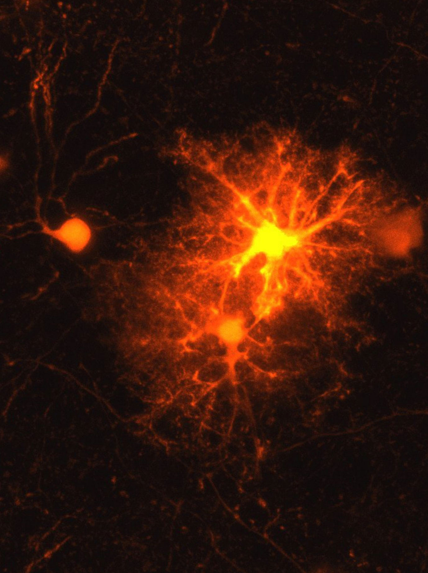
Ries began a systematic investigation using a mouse model, testing markers associated with every known cell type. Only one marker consistently responded. It corresponded to oligodendrocyte progenitor cells (OPCs). These precursor cells eventually develop into oligodendrocytes, which produce the myelin sheath surrounding axons. Axons are extensions that nerve cells use to communicate with each other.
The myelin sheath functions much like insulation around an electrical wire. In addition to supporting the transmission of signals, it also helps provide nutrients to nerve cells, making it essential for healthy brain function. In diseases such as multiple sclerosis (MS), this protective coating breaks down. Physical injuries can also damage the myelin layer, and in severe cases, neurons may die. For this reason, rebuilding the myelin sheath after injury is a critical part of brain repair.
Cells identified and a new mechanism discovered
Ries initially focused on describing the basic characteristics of these cells in his master’s thesis. “The topic remained so exciting that it became my doctoral thesis,” says the biologist. During this work, he demonstrated that OPCs multiply rapidly around the edges of injured brain tissue. Most of these cells then continue maturing and develop into oligodendrocytes that produce new myelin.
While studying these cells, Ries and his supervisor Deussing discovered an unexpected feature. In the region surrounding injuries, roughly one-third of OPCs began producing corticotropin-releasing hormone (CRH), a molecule that plays an important role in regulating the body’s stress response. Before this discovery, scientists did not know that OPCs were capable of producing neuropeptides such as CRH. The results of this research have been published in the journal Cell Reports.
The release of CRH occurs quickly. Scientists detected its presence within just a few hours after the injury occurred. Production then fades again after about three days. This short and rapid response suggests that the hormone may play an important role during the earliest stages of wound healing in the brain.
Researchers also found that one of the two known CRH receptors appears to be critical in this process. CRH receptor 1, which is present in a separate group of OPCs, responds to the CRH released nearby. When this receptor is absent, OPCs multiply more rapidly following injury. However, fewer of them ultimately mature into fully functional oligodendrocytes that remain in the tissue. These findings indicate that CRH helps control the timing of OPC development so that the myelin sheath can be properly restored.
What happens after birth?
OPCs are not only important after injuries. They also play a key role during normal brain development. Myelination, the process of building the myelin sheath around axons, occurs mainly after birth and continues until early adulthood.
Because CRH receptor 1 is present on OPCs even when there is no injury, the researchers wondered whether it might also influence myelination during normal brain maturation. To explore this question, they collaborated with additional scientists and examined myelination in several mouse models using different experimental methods.
Their observations showed that when CRH receptor 1 is missing, more OPCs are produced during early development. This shift has long-term effects on the structure of the brain. In adult mice lacking the receptor, researchers detected changes in myelination that were linked to thicker myelin sheaths, particularly around thinner axons.
These results suggest that CRH receptor 1 plays an important role in regulating myelination during brain development. In cases of injury, OPCs themselves release CRH in response to damage. During normal development, however, the scientists suspect that neurons supply the hormone instead. Their hypothesis is that developing neurons release CRH, which then influences how OPCs multiply and mature into oligodendrocytes that generate myelin.
Significance for psychiatric disorders
CRH is already known to be released by neurons during stressful conditions. Exposure to stress early in life is considered a significant risk factor for several psychiatric disorders.
“Our current findings suggest that in stress-associated psychiatric disorders such as depression, the CRH system in OPCs may play a greater role than previously known,” Deussing speculates.
In the long term, understanding this mechanism could open the door to new therapeutic strategies for mental health conditions.
Reference: “Neuropeptide CRH prevents premature differentiation of OPCs following CNS injury and in early postnatal development” by Clemens Ries, Tibor Stark, Benoit Boulat, Torben Ruhwedel, Jan Philipp Delling, Antonio Miralles Infante, Julia T. von Poblotzki, Alessandro Ulivi, Iven-Alex von Mücke-Heim, Simon Chang, Kenji Sakimura, Keiichi Itoi, Dennis B. Nestvogel, Alessio Attardo, Michael Czisch, Klaus-Armin Nave, Wiebke Möbius, Leda Dimou and Jan M. Deussing, 25 November 2025, Cell Reports.
DOI: 10.1016/j.celrep.2025.116474
Never miss a breakthrough: Join the SciTechDaily newsletter.
Follow us on Google and Google News.
1 Comment
I was under the impression that myelin sheaths are continuously stripped off and rebuilt, rather than just “one and done”. More research needed.