

The finding could eventually enable the use of DNA packaging in cells for disease diagnosis and treatment.
In preserved cells, newly developed fluorescent probes allowed researchers to pinpoint individual dye molecules to within 3 nanometers, close to the width of the DNA double helix itself. The same molecules can also reveal how DNA is packaged and moves inside living cells, giving scientists a way to examine chromatin at resolutions that were previously difficult to achieve without killing the cells.
When the team tested the probes on wax-preserved bowel tissue from three cancer patients, they found another striking difference. DNA in the tumors appeared noticeably looser and more spread out than in healthy tissue immediately beside it. Other research has found that DNA progressively unpacks as cancer develops, raising the possibility that its degree of folding could eventually provide information about how advanced or aggressive a tumor is.
Doctors currently assess these biopsies by eye using a staining technique that is more than a century old. The new approach, described in the journal Molecular Cell, could eventually add information about how DNA is arranged in three-dimensional space as another clue for cancer diagnosis and treatment.
“With the same dye we can do two very different things. In a living cell we can watch DNA moving, which tells us how chromatin, the natural state of DNA in cells, behaves. In a preserved cell we can zoom in until we are almost at the scale of the DNA molecule itself. Combining both approaches helps us see one of the main layers of control in human biology in unprecedented resolution,” explains ICREA Research Professor Pia Cosma, senior author of the study.
DNA folding controls which genes switch on
Each human cell packs about two meters (6.6 feet) of DNA into a tiny space. How tightly that DNA is folded helps determine which genes are active and which remain switched off.
Studying that organization in living cells has been difficult. Most images of DNA folding come from cells that are already dead because powerful microscopy techniques used to track individual components often depend on harsh chemicals and intense laser light that living cells cannot tolerate.
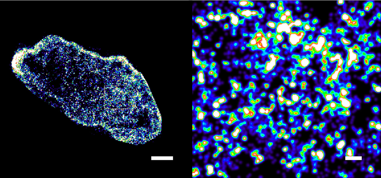
Blinking probes sharpen views of living DNA
Researchers at the Centre for Genomic Regulation (CRG) in Barcelona, the City University of Hong Kong and the Guangdong Provincial People’s Hospital (Guangdong Academy of Medical Sciences) Southern Medical University designed fluorescent probes that could overcome this limitation.
Known as HoTs, the dyes can enter living cells on their own and bind to DNA. They are engineered to blink intermittently, switching on and off rather than glowing continuously. The researchers tested them in living human skin cells and laboratory-grown HeLa cancer cells.
That blinking behavior is essential for producing sharper images. If all the dyes emitted light simultaneously, their signals would blur together. By detecting individual probes as they switch on, an advanced microscope can determine their locations. A computer program then combines thousands of snapshots to create an image about ten times sharper than one produced by a conventional microscope.
“The key challenge was to design fluorophores with the right blinking behavior for super-resolution imaging,” says Professor Hongyan Sun at City University of Hong Kong, co-senior author of the study.
A conventional microscope cannot clearly distinguish objects separated by less than about 200 nanometers. Using the new probes with a super-resolution technique called STORM, the researchers visualized DNA inside living cells at a resolution of 20 nanometers.
They achieved even greater precision in chemically preserved laboratory cells, where cellular structures are fixed in place. With another microscopy technique called MINFLUX, the team located individual dye molecules to within three billionths of a meter, or 3 nanometers.
“The diameter of the DNA double helix is approximately 2 nanometers, so 3 nanometer localization precision brings us remarkably close to the physical scale of the DNA molecule itself,” said Aiping Wang, first author of the study.
The method reveals differences beyond cancer
The probes also worked outside human cells. Researchers applied them to slices of zebrafish eye tissue. Zebrafish can regenerate damaged retina, a process thought to involve cells loosening their DNA so they can become flexible again.
The researchers also tested whether artificial intelligence could distinguish cells based on these detailed images. They used AINU, short for “AI of the Nucleus,” a program the team introduced in 2024 that examines super-resolution images of nuclear DNA for patterns too subtle for the human eye.
After training on the new images of living cells, AINU distinguished skin cells from stem cells with 96 to 98 percent accuracy. Although the two cell types contain identical DNA, they package it differently, and the AI was able to detect those differences.
AINU originally worked only with cells that had been killed and preserved. Combining it with the new fluorescent probes now allows the program to analyze living cells. The researchers hope the same principle could eventually help distinguish cancerous tissue from healthy tissue or identify promising stem cells for regenerative medicine.
The sharpest images still require preserved cells
The technique still has important limitations. The fluorescent molecules coat DNA broadly rather than identifying a specific gene, and the highest-resolution images were obtained from preserved rather than living cells.
The team is now pursuing follow-up work on this limitation. MINFLUX also has the potential to image the probes in living cells.
Reference: “HoT auto-blinking probes enable real-time, super-resolution chromatin imaging in live cells and tissues” by Aiping Wang, Shanshan Liu, Heng Shi, Mikhail Rotkevich, Carlotta Viana, Hui Zhong, Limei Zhong, Lingkuan Meng, Yanwu Li, Guirong Chen, Xiaoming Zhu, Zhenzhen Ma, Bing Fu, Alvaro Castells-Garcia, Jie Zhang, Hongyan Sun and Maria Pia Cosma, 28 August 2026, Molecular Cell.
DOI: 10.1016/j.molcel.2026.08.010
Never miss a breakthrough: Join the SciTechDaily newsletter.
Follow us on Google and Google News.