Scientists apply principles of math and physics to unravel the mystery of how the endoplasmic reticulum, an organelle vital to cellular life, constantly reshapes and reorganizes itself.
As a second-year Ph.D. student and physicist, Zuben Scott hadn’t thought much about the endoplasmic reticulum since learning about cell structures as a high school freshman. Then a potential graduate adviser, Elena Koslover, suggested he study it. She showed him images and videos captured under a microscope that revealed an intricate mesh.
“It was very beautiful,” he says of the organelle better known as simply the ER. “It was shocking to me to see how this complex network could form within cells.”
Scott was equally intrigued by the question posed by Koslover, an associate professor of physics at the University of California, San Diego. Although best known as the site where proteins are assembled and prepared for their functions, the ER does much more. For example, it produces certain hormones and components of the cell membrane, and stores calcium ions, which cells use to coordinate responses to stimuli.
These molecules move through the ER’s elaborate structure, and Koslover, who studies transport in cells from a physics perspective, wanted to investigate how. To do so, they need to account for the ever-changing nature of this organelle.
“Constantly, every minute, the ER is restructuring and shifting around,” Koslover says.
Working with the same images, which were taken by Koslover’s collaborator Laura Westrate, Scott eventually devised a model to describe this continuous reconfiguration. This research, published recently in Proceedings of the National Academy of Sciences, uncovers the unique dynamics governing the ER’s evolution and addresses the long-standing mystery of just how this organelle sustains life at the cellular level, with implications for understanding disease.
A new kind of cellular network
Before he took on the ER, Scott, who has since joined Adrian Jacobo’s research group at the Chan Zuckerberg Biohub San Francisco as a scientist, had dabbled in biophysics. The summer before his senior year as a physics major at Reed College, he first encountered this interdisciplinary field through a data-analysis internship working on a super-resolution imaging technique, with Xiaolin Nan at Oregon Health & Science University.
The experience piqued his interest, but also revealed how much biology he had to learn to complement his knowledge of physics. “Xiaolin told me his seven-year-old knew more biology than me,” Scott says. “But eventually, I got there.”
In Koslover’s lab, Scott became the designated “ER person.” For his project, he focused on the tube-filled section of the organelle adjacent to the cell’s membrane known as the peripheral ER. (Another portion, composed of sheets stacked like the levels of a parking garage, enfolds the nucleus.) He approached the tubular ER as a network, a term in physics that describes a set of connected points — think users on a social media platform, the intersections of a city’s roads, or in the case of the ER, the junctions where its tiny tubes meet, three at a time.
Cells contain other networks too. Scientists describe the cell’s internal skeleton and its energy-converting mitochondria this way. But the dynamics that define other cellular networks don’t apply to the peripheral ER.
“The junctions actually slide,” says Greg Huber, a UC San Francisco biophysicist and coauthor on the PNAS paper, describing how the three-tube connection points respond to the forces transmitted through the network of tubules. But this movement resembles that of a liquid, not a solid, so “the physical material that makes up a junction at one time will contain different molecules at a later time.”
Huber, who previously led Biohub SF’s Physical Biology and Biophysical Theory Group, had been working on his own model for the peripheral ER when he joined Scott and Koslover’s project. To describe its behavior, he had taken to calling it a “liquid network,” but notes that, unlike an everyday liquid, the tubular ER generates its own shape. It is its own container, he says.
Simple model, complicated structure
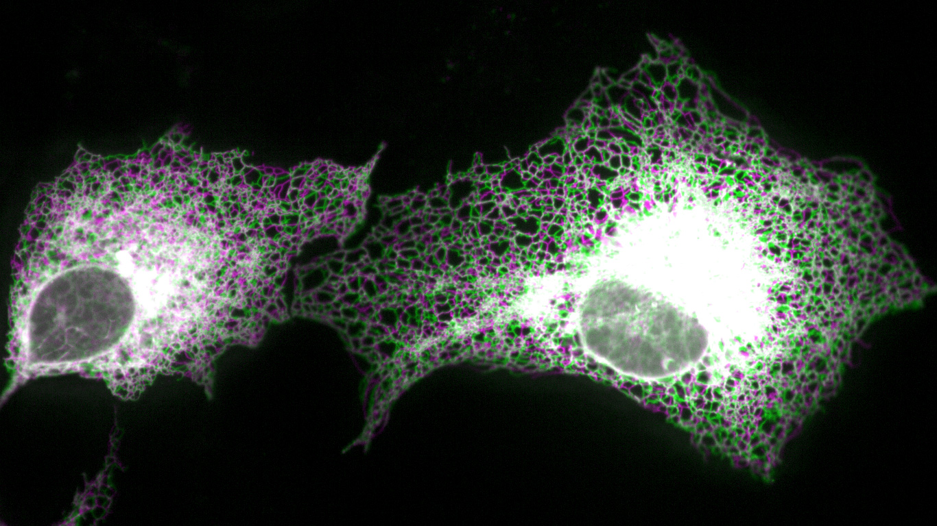
To visualize this dynamic network of tubules, Westrate, of Calvin University, took advantage of a property of cells from a line known as COS-7: when grown in culture, these cells spread out like fried eggs. Thick in the middle but thin at the edges, this distinctive shape squishes the peripheral ER into nearly two dimensions, simplifying the task of studying it with imaging tools.
The time-lapse images Westrate captured show new tubules spontaneously branching from and connecting to the existing network or other parts of the cell. Simultaneously, tension causes junctions to slide, shrinking tubules and closing the rings they form — leading to a network of simultaneously growing and shrinking shapes.
Scott and Samuel Steen, from Westrate’s group, defined these dynamics mathematically by counting new tubules and determining the average area of the polygons the tubules enclosed. With these measurements, they derived the model’s two parameters: tubule growth rate and the mobility of the junctions. Using these parameters, it predicts the structural features of the ER with no further fiddling.
“The best part about this model,” Scott says, “is its simplicity.”
The same rules would likely apply to a 3D model of the ER; however, a 3D model would also need to account for the tubules’ widths, and this measurement would determine whether a growing tube intersects an existing one, or passes it by, he says. But even in 2D, this mathematical representation helps explain how the ER can, as previous research has shown, move about to explore most of a cell’s interior, delivering proteins, calcium, and other molecules as needed.
In their paper, the team calls the peripheral ER an “active liquid network,” a term Huber coined, to capture not only the liquid-like sliding of the junctions but also the growth of new tubules from existing ones.
“We have contributed — I hate to say it because the term’s so overused — a new paradigm,” Huber says, and he suspects that there are other examples of active liquid networks within the specialized internal structures of cells.
By offering insight on how cells function normally, the team’s model is relevant to understanding how things go awry in disease, particularly Alzheimer’s disease, amyotrophic lateral sclerosis, and spastic paraplegia, which studies have linked to changes in the ER’s shape. Their research could also have more general implications for ER dysfunction in numerous other conditions, including heart disease and diabetes.
Exploring life through physics
When Scott joined Jacobo’s Quantitative Tissue Morphogenesis group in October, he traded the ER for the neuromast, a sensory organ found on the sides of the zebrafish that the lab studies to explore the dynamics involved in arranging cells to form organs during development.
He is also planning a collaboration with colleagues at Stanford to examine the processes that maintain the midsection of the tube that forms the gut in fruit flies. The zebrafish neuromast and Drosophila gut have little in common with each other, let alone the ER, but Scott believes physical principles can contribute to understanding how these diverse structures form.
After his time immersed in the ER and as a new member of an experimental biology lab, Scott now describes himself as “slightly less inexperienced” in biology, a field in which he still feels somewhat like an outsider.
“I constantly oscillate between states of taking biology for granted and being in awe of the complexity of living systems,” he says.
Reference: “The endoplasmic reticulum as an active liquid network” by Zubenelgenubi C. Scott, Samuel B. Steen, Greg Huber, Laura M. Westrate and Elena F. Koslover, 11 October 2024, Proceedings of the National Academy of Sciences.
DOI: 10.1073/pnas.2409755121
Never miss a breakthrough: Join the SciTechDaily newsletter.
Follow us on Google and Google News.