
New technique could be used in medical diagnostics and advanced manufacturing.
Ever since Antonie van Leeuwenhoek discovered the world of bacteria through a microscope in the late seventeenth century, humans have tried to look deeper into the world of the infinitesimally small.
There are, however, physical limits to how closely we can examine an object using traditional optical methods. This is known as the ‘diffraction limit’ and is determined by the fact that light manifests as a wave. It means a focused image can never be smaller than half the wavelength of light used to observe an object.
Attempts to break this limit with “super lenses” have all hit the hurdle of extreme visual losses, making the lenses opaque. Now physicists at the University of Sydney have shown a new pathway to achieve superlensing with minimal losses, breaking through the diffraction limit by a factor of nearly four times. The key to their success was to remove the super lens altogether.
The research is published today (October 18) in the journal Nature Communications.
Implications for Science and Beyond
The work should allow scientists to further improve super-resolution microscopy, the researchers say. It could advance imaging in fields as varied as cancer diagnostics, medical imaging, or archaeology and forensics.
Lead author of the research, Dr. Alessandro Tuniz from the School of Physics and University of Sydney Nano Institute, said: “We have now developed a practical way to implement superlensing, without a super lens.
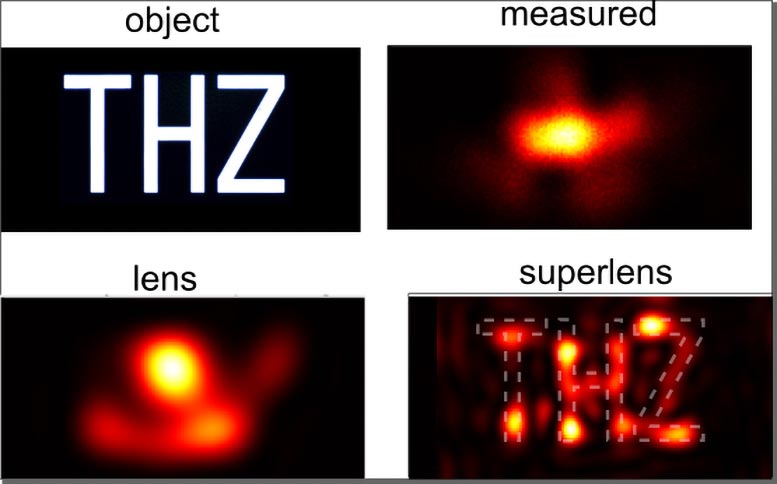
“To do this, we placed our light probe far away from the object and collected both high- and low-resolution information. By measuring further away, the probe doesn’t interfere with the high-resolution data, a feature of previous methods.”
Previous attempts have tried to make super lenses using novel materials. However, most materials absorb too much light to make the super lens useful.
Dr. Tuniz said: “We overcome this by performing the superlens operation as a post-processing step on a computer, after the measurement itself. This produces a ‘truthful’ image of the object through the selective amplification of evanescent, or vanishing, light waves.
Practical Applications and Future Prospects
Co-author, Associate Professor Boris Kuhlmey, also from the School of Physics and Sydney Nano, said: “Our method could be applied to determine moisture content in leaves with greater resolution, or be useful in advanced microfabrication techniques, such as non-destructive assessment of microchip integrity.
“And the method could even be used to reveal hidden layers in artwork, perhaps proving useful in uncovering art forgery or hidden works.”
Typically, superlensing attempts have tried to home in closely on the high-resolution information. That is because this useful data decays exponentially with distance and is quickly overwhelmed by low-resolution data, which doesn’t decay so quickly. However, moving the probe so close to an object distorts the image.
“By moving our probe further away we can maintain the integrity of the high-resolution information and use a post-observation technique to filter out the low-resolution data,” Associate Professor Kuhlmey said.
The research was done using light at terahertz frequency at millimeter wavelength, in the region of the spectrum between visible and microwave.
Associate Professor Kuhlmey said: “This is a very difficult frequency range to work with, but a very interesting one, because at this range we could obtain important information about biological samples, such as protein structure, hydration dynamics, or for use in cancer imaging.”
Dr. Tuniz said: “This technique is a first step in allowing high-resolution images while staying at a safe distance from the object without distorting what you see.
“Our technique could be used at other frequency ranges. We expect anyone performing high-resolution optical microscopy will find this technique of interest.”
Reference: “Subwavelength terahertz imaging via virtual superlensing in the radiating near field” by Alessandro Tuniz, and Boris T. Kuhlmey, 18 October 2023, Nature Communications.
DOI: 10.1038/s41467-023-41949-5
Funding: Australian Research Council
Never miss a breakthrough: Join the SciTechDaily newsletter.
Follow us on Google and Google News.