Close Menu
    Facebook X (Twitter) Instagram
    SciTechDaily
    • Biology
    • Chemistry
    • Earth
    • Health
    • Physics
    • Science
    • Space
    • Technology
    Facebook X (Twitter) Pinterest YouTube RSS
    SciTechDaily
    Home»Technology»Transforming Nanofiber Membranes Into Complex 3D Shapes for Biomedical Applications
    Technology

    Transforming Nanofiber Membranes Into Complex 3D Shapes for Biomedical Applications

    By SciTechDailyMay 12, 2020No Comments3 Mins Read
    Facebook Twitter Pinterest Telegram LinkedIn WhatsApp Email Reddit
    Share
    Facebook Twitter LinkedIn Pinterest Telegram Email Reddit
    Cylinder-Shaped Nanofiber Scaffold Schematic
    Schematic illustrating the procedure of converting a 2D nanofiber mat into a cylinder-shaped nanofiber scaffold with (a) a hollow tube-shaped mold for a cylindrical shape and (b) a customized scaffold for a tube shape. The photographs (c) represent the different scaffold that can be created with different mold shapes. Credit: Jingwei Xie

    Researchers develop a new way to create 3D scaffolds for biomedical applications.

    In the movie “Transformers,” cars morph into robots, jets, or a variety of machinery. A similar concept inspired a group of researchers to combine gas foaming, which is a blend of chemicals that induces gas bubbling, and 3D molding technologies to quickly transform electrospun membranes into complex 3D shapes for biomedical applications.

    In Applied Physics Reviews, from AIP Publishing, the group reports on its new approach that demonstrates significant improvements in speed and quality compared with other methods. The work is also the first successful demonstration of formation of 3D neural tissue constructs with an ordered structure through differentiation of human neural progenitor/stem cells on these transformed 3D nanofiber scaffolds.

    “Electrospinning is a technology to produce nanofiber membranes,” said co-author Jingwei Xie, at the University of Nebraska Medical Center. “The physics principle behind it involves applying an electrical force to overcome the surface tension of a solution to elongate a solution jet into continuous and ultrafine fibers after solvent evaporation.”

    Due to an intrinsic property of electrospinning, nanofibers are often deposited to form 2D membranes or sheets with dense structures and small pore sizes that are less than the size of cells.

    “This greatly inhibits the applications of electrospun nanofibers, because cells fail to seed or penetrate throughout the nanofiber membranes, which is undesirable,” he explained.

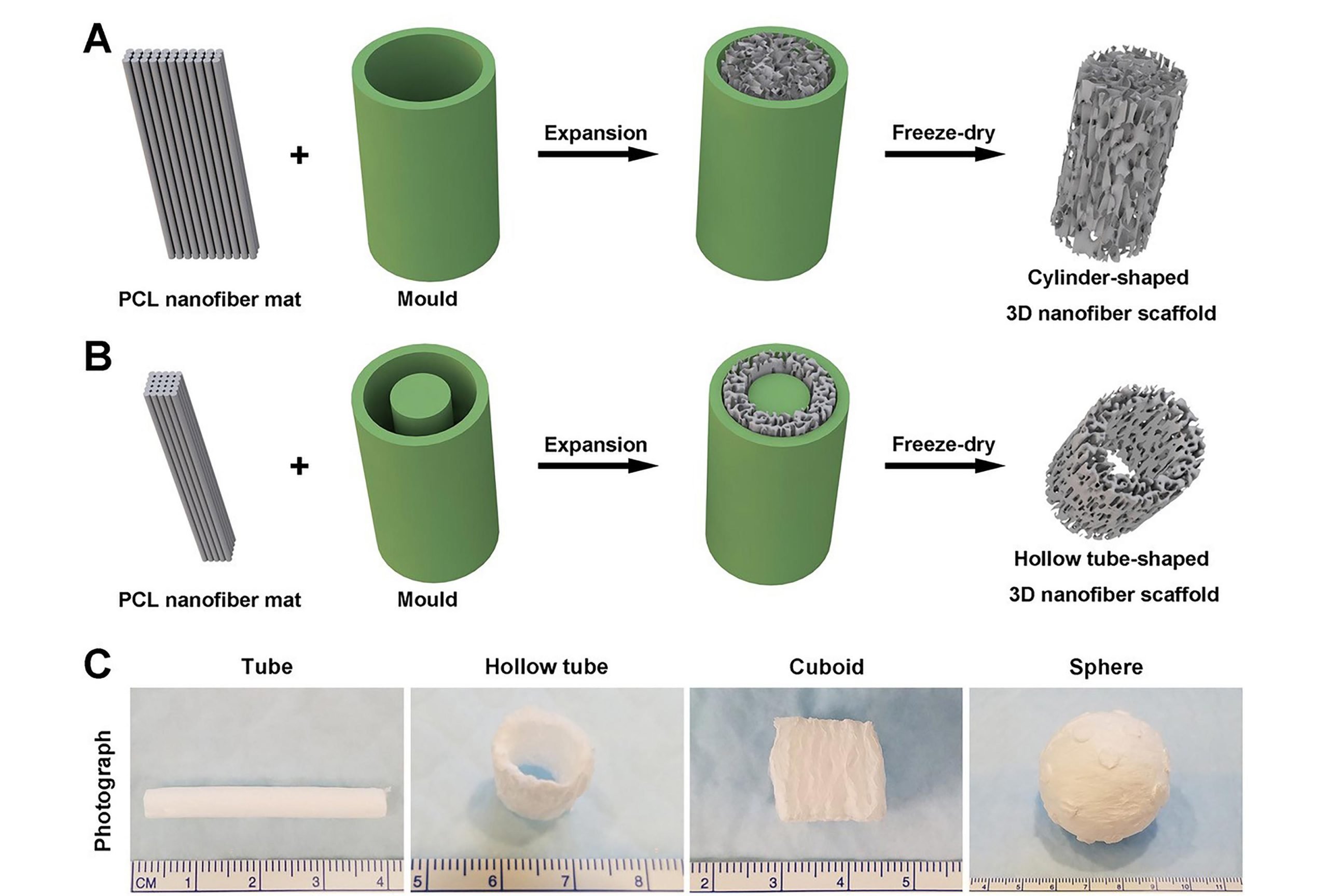
    Researchers combined gas foaming and 3D molding concepts to expand nanofiber membranes within a confined space to form predesigned 3D nanofiber objects in cylindrical, cuboid, spherical, and irregular shapes.

    “Our 3D objects have the appropriate pore size and controlled fiber alignment for guiding and enhancing cell penetration to form new tissue,” Xie said.

    The group’s work is significant because it can be done within an hour. Other methods can require up to 12 hours to complete the transformation process.

    “Thanks to the ability to mimic the architecture of extracellular matrix, electrospun nanofibers show great potential in applications such as tissue engineering, regenerative medicine and tissue modeling,” said Xie.

    One of the group’s most intriguing findings is that after coating 3D nanofiber objects with gelatin, they exhibit superelasticity and shape recovery.

    “Gelatin-coated, cube-shaped scaffolds functionalized with polypyrrole coatings exhibited dynamic electrical conductivity during cyclical compression,” he said.

    They also demonstrated that cuboid-shaped nanofiber objects were effective for compressible hemorrhage in a pig liver injury model.

    In the future, the group’s method may help “enable therapeutic-free biomaterials for tissue repair and regeneration, such as using predesigned nanofiber objects to fit irregular tissue defects,” Xie said. “Beyond that, superelasticity and shape recovery could allow 3D-nanofiber objects to be applied in a minimally invasive manner.”

    Reference: “Fast transformation of 2D nanofiber membranes into pre-molded 3D scaffolds with biomimetic and oriented porous structure for biomedical applications” by Sixuan Chen, Johnson V. John, Alec McCarthy, Mark A. Carlson, Xiaowei Li and Jingwei Xie, 12 May 2020, Applied Physics Reviews.
    DOI: 10.1063/1.5144808

    Never miss a breakthrough: Join the SciTechDaily newsletter.
    Follow us on Google and Google News.

    American Institute of Physics Biomedical Engineering Biotechnology Cell Biology Nanotechnology
    Share. Facebook Twitter Pinterest LinkedIn Email Reddit

    Related Articles

    COVID-19 Coronavirus Breathalyzer Test Developed

    DARPA Invisible Man: Human Cells Engineered With Squid-Like Transparency

    Discovery of Complex Nanometric Geometric Shapes on Dragonfly Wings Improves the Efficiency of Antibacterial Surfaces

    Nanodevices Track Cells From the Inside, Show How They Develop With Time

    Bionic Tactile Enhancement – Highly Sensitive Sensors Inspired by Spiders Improve Sense of Touch

    New Nanofiber Dressings Dramatically Accelerate Healing and Tissue Regeneration

    Single-Stranded DNA and RNA Origami That Can Autonomously Fold Into Defined Structures

    Miniature System Can Encapsulate and Analyze Biomarkers in Sweat

    New Technique Enlarges Tissue Samples, Making Them Easier to Image

    Leave A Reply Cancel Reply

    • Facebook
    • Twitter
    • Pinterest
    • YouTube

    Don't Miss a Discovery

    Subscribe for the Latest in Science & Tech!

    Trending News

    The Universe Is Expanding Too Fast and Scientists Can’t Explain Why

    “Like Liquid Metal”: Scientists Create Strange Shape-Shifting Material

    Early Warning Signals of Esophageal Cancer May Be Hiding in Plain Sight

    Common Blood Pressure Drug Shows Surprising Power Against Deadly Antibiotic-Resistant Superbug

    Scientists Uncover Dangerous Connection Between Serotonin and Heart Valve Disease

    Scientists Discover a “Protector” Protein That Could Help Reverse Hair Loss

    Bone-Strengthening Discovery Could Reverse Osteoporosis

    Scientists Uncover Hidden Trigger Behind Stem Cell Aging

    Follow SciTechDaily
    • Facebook
    • Twitter
    • YouTube
    • Pinterest
    • Newsletter
    • RSS
    SciTech News
    • Biology News
    • Chemistry News
    • Earth News
    • Health News
    • Physics News
    • Science News
    • Space News
    • Technology News
    Recent Posts
    • Millions Take These IBS Drugs, But a New Study Finds Serious Risks
    • 5 Common Myths About Learning a New Language, Debunked
    • The Neanderthal “Love Story” Isn’t What It Seems
    • Scientists Unlock Hidden Secrets of 2,300-Year-Old Mummies Using Cutting-Edge CT Scanner
    • Men vs. Women: Scientists Uncover Dramatic Differences in How the Immune System Ages
    Copyright © 1998 - 2026 SciTechDaily. All Rights Reserved.
    • Science News
    • About
    • Contact
    • Editorial Board
    • Privacy Policy
    • Terms of Use

    Type above and press Enter to search. Press Esc to cancel.