Recent research identifies DNA methylation’s critical role in craniofacial development, providing a pathway toward preventing cleft lip and palate by understanding environmental impacts on genetic expression.
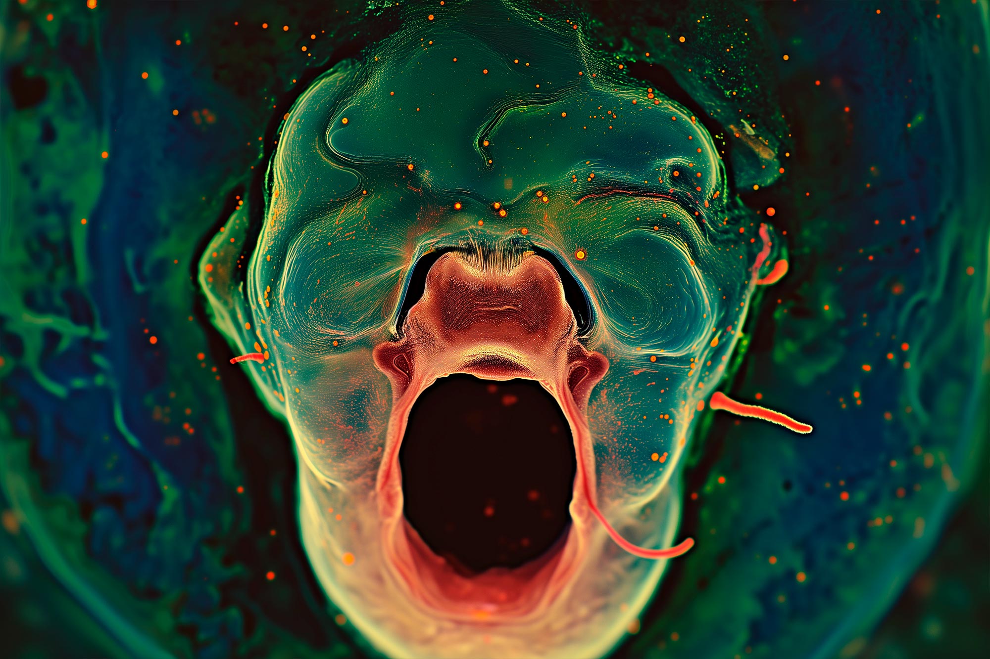
Cleft lip and palate are the most common craniofacial birth defects in humans, affecting more than 175,000 newborns around the world each year. Yet despite decades of research, it’s still not known what causes most cases or what can be done to prevent them. But a recent study from the University of Wisconsin School of Veterinary Medicine (SVM) has uncovered new information about orofacial development in mice that researchers believe could one day help reduce the risk of these birth defects in humans.
DNA Methylation and Craniofacial Development
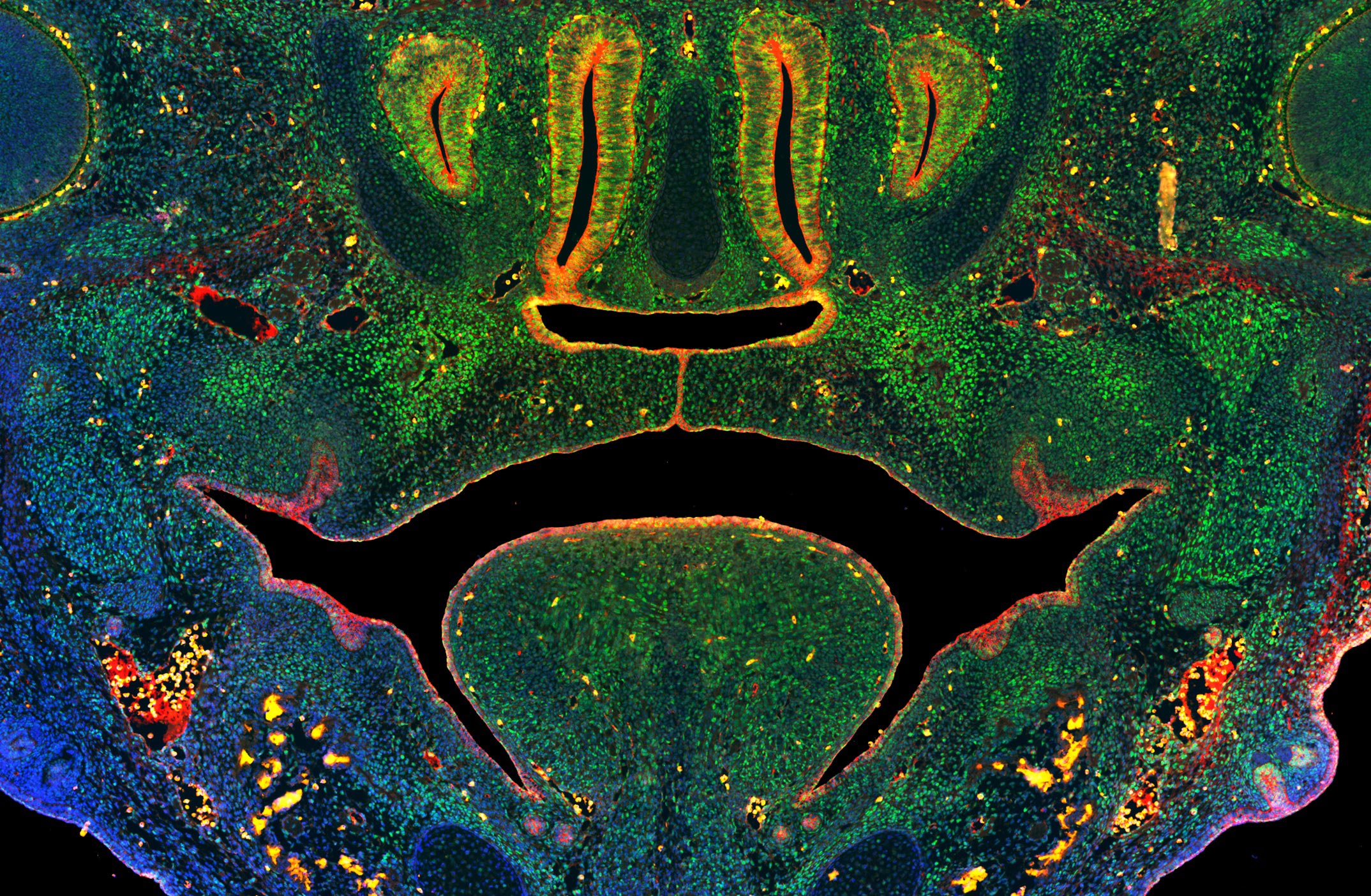
Published this week in the Proceedings of the National Academy of Sciences (PNAS) the study provides the first direct evidence of a mechanism called DNA methylation being required for craniofacial development. DNA methylation is a process where a group of molecules are added to DNA that change the expression of genes without actually altering the DNA sequence. It’s also affected by various environmental factors. The researchers discovered that disruption to DNA methylation interferes with development of the lip and palate and causes these birth defects in mice.
Led by Robert Lipinski, associate professor of comparative biosciences at the UW School of Veterinary Medicine, the research is an important step toward developing preventive strategies that could one day lessen the risk of cleft lip and palate, known collectively as orofacial clefts (OFCs), in both animals and humans.
Environmental Influences on Orofacial Clefts
“We knew from past research that genetics and the environment interact to cause these types of birth defects, but our understanding of the environmental component lagged far behind that of genetics.” says Lipinski. “Unlike genetics, we don’t have a permanent record of the prenatal environment that can be examined retrospectively, but connecting OFCs to DNA methylation helps narrow our focus on the particular environmental influences that modify the risk for these types of birth defects.”
His team’s work confirmed the essential role of DNA methylation in regulating orofacial development during embryonic development and demonstrates how disruptions to that process alter the ability of stem cells to form the connective tissue of craniofacial bone and cartilage, resulting in OFCs.
Lipinski and his team arrived at these results by first genetically manipulating DNA methylation in two separate groups of mouse embryos. The experiments resulted in seemingly contradictory results, with OFCs developing in one group of mice, but not the other. To understand why there was a difference between the groups, the team conducted another round of experiments in which they inhibited DNA methylation in mouse embryos at different stages of development. The timing of when DNA methylation occurs, was critical to the development of orofacial clefts.
They found that exposure on the 10th gestational day resulted in OFCs but administering the same inhibitor just 48 hours later resulted in normal orofacial development.
Narrowing the Research Focus
Identifying this narrow window of gestational sensitivity is important, Lipinski says, because it not only helps narrow the focus of the next stage of his team’s research but it will also help design future public education initiatives once more is known about the modifiable environmental and behavioral risk factors that impact OFC risk in humans. The 10th gestational day in mouse embryos corresponds with the beginning of the 5th week of embryonic development in humans–a stage at which many pregnancies may not yet be recognized.
“We know DNA methylation can be influenced by a variety of environmental factors, including maternal stress, diet, and exposure to drugs, toxins and environmental pollutants, and having a better understanding how orofacial development is regulated by environmentally sensitive mechanisms could directly inform birth defect prevention strategies,” he says. “This next phase of our team’s research is focused on identifying specific factors that influence DNA methylation during orofacial development and which could therefore alter OFC risk.”
Advancing Research with New Models
Lipinski and his team are uniquely positioned to pursue this next stage of research because of another important outcome of the study: a new in vitro model the team developed. The model will allow them to rapidly screen thousands of dietary and environmental factors in a laboratory dish before testing the impact of specific factors on cleft susceptibility in mouse models.
The results in cell and animal models will help the researchers more quickly and accurately identify factors likely to be of consequence to human development.
The Impact of Orofacial Clefts
Orofacial clefts of the upper lip and palate affect approximately 1 in 700 newborns, and individuals with OFCs navigate feeding difficulties as infants that require multiple surgeries, dental procedures, and speech therapy during childhood and adolescence. Studies have shown higher mortality rates at all stages of life for individuals with OFCs.
Reference: “Disruption of DNA methylation–mediated cranial neural crest proliferation and differentiation causes orofacial clefts in mice” by Caden M. Ulschmid, Miranda R. Sun, Christopher R. Jabbarpour, Austin C. Steward, Kenneth S. Rivera-González, Jocelyn Cao, Alexander A. Martin, Macy Barnes, Lorena Wicklund, Andy Madrid, Ligia A. Papale, Diya B. Joseph, Chad M. Vezina, Reid S. Alisch and Robert J. Lipinski, 9 January 2024, Proceedings of the National Academy of Sciences.
DOI: 10.1073/pnas.2317668121
This study was supported by funding from the National Institutes of Health under award numbers R03DE027162, R56DE030917, RO1DE032710, U01 DK11807, and R01DK099328, and T32ES007015. Additional support was also provided by the University of Wisconsin Hilldale Undergraduate Research Award.
Never miss a breakthrough: Join the SciTechDaily newsletter.
Follow us on Google and Google News.
2 Comments
This approach of narrowing the focus is going to have some of the greatest steps forward in the full understanding of birth defects possible causes pollutants or environmental down to hereditarily transmitted causes. Maybe the mechanisms of how a process starts and progresses will be more defined.
Based in part on family and personal history, I’ve already written a few of my now senior lay findings of connections between allergies, maternal calcium deficiency and cleft lips and/or palates. During my lay investigations of Dr. Arthur F. Coca’s (by 1934; my) kind of very, very mild non-IgE-mediated food allergies aggravated (or not) with FDA approved food poisoning (namely added cultured “free” MSG and soy processed mostly with toxic hexane) and/or related/resultant medical errors I discovered my own deciduous bicuspids are related to cleft lips and palates. Since, I postulate that food rationing during W.W.II and/or my mother’s own allergy to milk were/are the primary reasons, with standard blood serum testing for calcium being unreliable due to blood pH regulation. I hope this helps you narrow your focus.