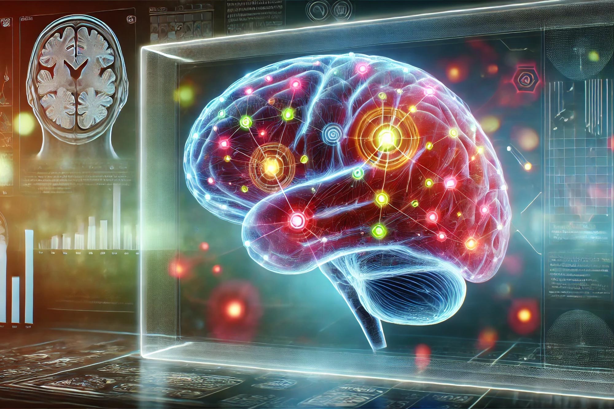
Researchers have developed a novel method for delivering fluorescent sensors across the blood-brain barrier (BBB) to monitor neurotransmitter levels in the brain, which could significantly advance Alzheimer’s disease diagnosis and treatment.
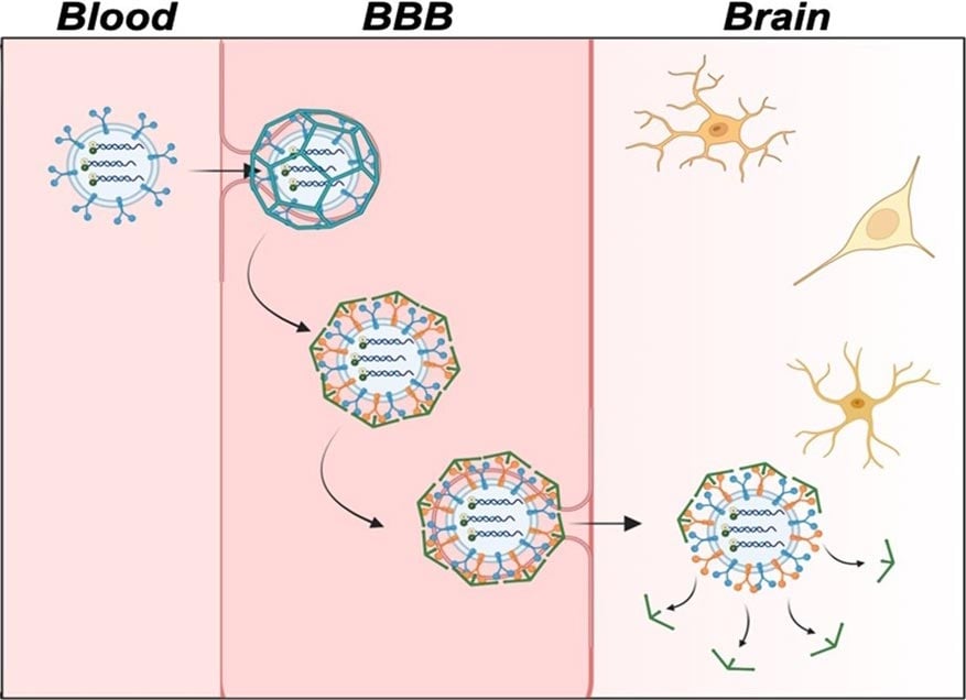
The method involves encapsulating ATP aptamer sensors in exosomes, which are able to cross the BBB and provide real-time imaging of neurotransmitter levels. Initial tests in mice have shown promising results, with sensors accumulating in the brain and identifying low ATP levels in areas affected by Alzheimer’s.
Innovative Approach to Brain Imaging
Neurotransmitter levels in the brain can indicate brain health and neurodegenerative diseases like Alzheimer’s. However, the protective blood-brain barrier (BBB) makes delivering fluorescent sensors that can detect these small molecules to the brain difficult. Now, researchers in ACS Central Science demonstrate a way of packaging these sensors for easy passage across the BBB in mice, allowing for improved brain imaging. With further development, the technology could help advance Alzheimer’s disease diagnosis and treatment.
It is common for neurotransmitter levels to decrease with age, but low levels of the neurotransmitter adenosine triphosphate (ATP) can be an indication of Alzheimer’s disease. To measure the location and amount of ATP in the brain, researchers have developed fluorescent sensors from pieces of DNA called aptamers that light up when they bind to a target molecule. Methods for delivering these sensors from the bloodstream to the brain have been developed, but most contain synthetic components that can’t easily cross the BBB. To develop sensors for live brain imaging, Yi Lu and colleagues encapsulated an ATP aptamer sensor in brain-cell-derived microscopic vesicles called exosomes. They tested the new sensor delivery system in lab models of the BBB and in mouse models of Alzheimer’s disease.
Successful Sensor Delivery Using Exosomes
The BBB laboratory model consisted of a layer of endothelial cells on top of a solution containing brain cells. The researchers’ sensor-loaded exosomes were nearly four times more efficient than conventional sensor delivery systems at passing through the endothelial barrier and releasing the fluorescent sensor into the brain cells. This was confirmed by measuring the observed level of ATP-binding-induced fluorescence. Next, Lu’s team injected mouse models of Alzheimer’s disease with either the sensor-loaded exosomes or free-floating unloaded sensors. By measuring fluorescence signals in the mice, the researchers found that the free-floating sensors stayed mainly in the blood, liver, kidneys, and lungs, while sensors delivered via exosomes accumulated in the brain.
Promising Results in Alzheimer’s Mouse Models
In mouse models of Alzheimer’s disease, the exosome-delivered sensors identified the location and concentration of ATP in different regions of the brain. Specifically, they observed low levels of ATP in the hippocampus, cortex, and subiculum regions of the brain, which are indicative of the disease. The researchers say that their exosome-loaded ATP-reactive sensors show promise for non-invasive live brain imaging and could be developed further to create sensors for a range of clinically relevant neurotransmitters.
Reference: “Delivering DNA Aptamers Across the Blood–Brain Barrier Reveals Heterogeneous Decreased ATP in Different Brain Regions of Alzheimer’s Disease Mouse Models” by Mandira Banik, Aaron P. Ledray, Yuting Wu and Yi Lu, 31 July 2024, ACS Central Science.
DOI: 10.1021/acscentsci.4c00563
The authors acknowledge funding from the U.S. National Institutes of Health (NIH), Welch Foundation, NIH Chemistry-Biology Interface Training Program at the University of Illinois Urbana-Champaign and a National Science Foundation Graduate Research Fellowship.
Never miss a breakthrough: Join the SciTechDaily newsletter.
Follow us on Google and Google News.