A new study reveals that depression suppresses microglial activity, affecting brain plasticity. Targeting microglial function may offer a novel approach for future antidepressants.
A recent research study from the Netherlands Institute for Neuroscience has discovered that individuals suffering from depression have fewer microglial cells. What does that mean?
Depression is a serious condition that greatly contributes to the overall global disease burden. It’s also a leading cause of disability on a worldwide scale. Given that up to 30% of patients experience resistance to available treatments, there is an urgent need for more understanding of the disease’s underlying mechanisms and for the development of new therapeutic approaches.
Previous research showed that patients with depression have altered levels of inflammatory markers. In addition, depression has been linked to chronic inflammatory diseases, such as rheumatism, inflammatory bowel disease, and multiple sclerosis. These results suggest that inflammation of the brain may play a role in depression. But is that true?
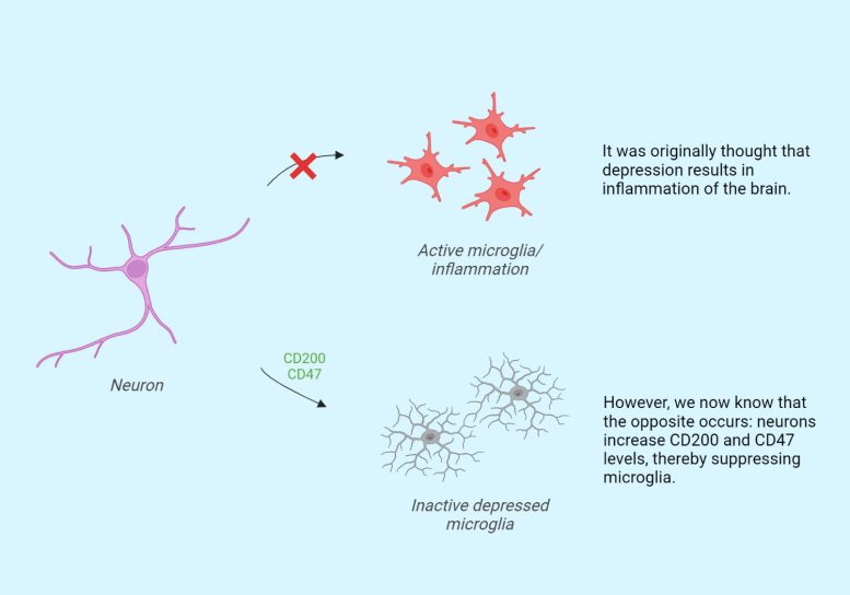
A new study by Karel Scheepstra and his team, supervised by Inge Huitinga and Jörg Hamann, looked at post-mortem human brain tissue from people with depression. This brain tissue comes from recently deceased donors who donated their brains to the Dutch Brain Bank for Psychiatry (NHB-Psy). And what did they find? A certain type of immune cells in our brain, called microglial cells, are less active in people with depression. Contrary to expectations, the opposite of inflammation actually occurs: the immune cells are suppressed.
Neurons Affect Microglia
Microglial cells are important because they maintain contact points between neurons (synapses), thereby helping neurons communicate efficiently with each other. In addition, microglial cells constantly scan the central nervous system for damaged neurons, synapses, and pathogens. In the samples from people with depression, only microglial cells near neurons showed reduced activity. The team, therefore, investigated whether neurons send signals to the microglial cells during depression, making them less active. And this indeed turned out to be the case.
Karel Scheepstra (researcher involved in the study and also working as a psychiatrist at Amsterdam UMC): ‘During the study, we used fresh tissue immediately after death to isolate microglia and compared these between depressed people and controls. We saw abnormal microglia in depressed patients, with the greatest abnormalities seen in patients who were most depressed just before death. Interestingly, abnormalities were only seen in the gray matter and not in the white matter of the brain. This suggests that there is a likely interaction between the microglia and the structures located in the gray matter: the neurons and synapses.”
“We also looked at the type of alterations. We’ve hypothesized for years that depression is associated with inflammation of the brain, but we’re now seeing just the opposite: not neuroinflammation, but rather an immune-suppressed type of microglia. We termed them ‘depressed microglia’ and we wondered how exactly this is possible. The proteins CD200 and CD47 are located on brain cells and synapses. They interact with microglia and are, as it were, a kind of ‘don’t eat me signal’. What we saw is that these proteins were elevated, resulting in suppressed microglia, thereby possibly preventing them from clearing damaged connections.”
Neuroplasticity
“Depression is thought to have something to do with a change in neuroplasticity: the ability to make new connections between neurons. A relatively new antidepressant is esketamine, a drug that intervenes in this process and ensures that more connections start to grow again. In this study, we show that there is a disturbed neuron-microglia interaction. The next step would be to see what exactly the consequences of the inactive microglia are for the maintenance and formation of connections between neurons.’
‘If we know where things go wrong in the process, this can provide targets for new medication. Can we make these microglia more active again? And what effect does this have on the course of the disease? For now, we have shown that the brains of people who were depressed during life show altered cell activity. This gives us a better understanding of what goes wrong, which we can then build on.”
Reference: “Microglia transcriptional profiling in major depressive disorder shows inhibition of cortical grey matter microglia” by Karel W.F. Scheepstra, Mark R. Mizee, Jackelien van Scheppingen, Adelia Adelia, Dennis D. Wever, Matthew R.J. Mason, Marissa L. Dubbelaar, Cheng-Chih Hsiao, Bart J.L. Eggen, Jörg Hamann and Inge Huitinga, 28 April 2023, Biological Psychiatry.
DOI: 10.1016/j.biopsych.2023.04.020
Never miss a breakthrough: Join the SciTechDaily newsletter.
Follow us on Google and Google News.