
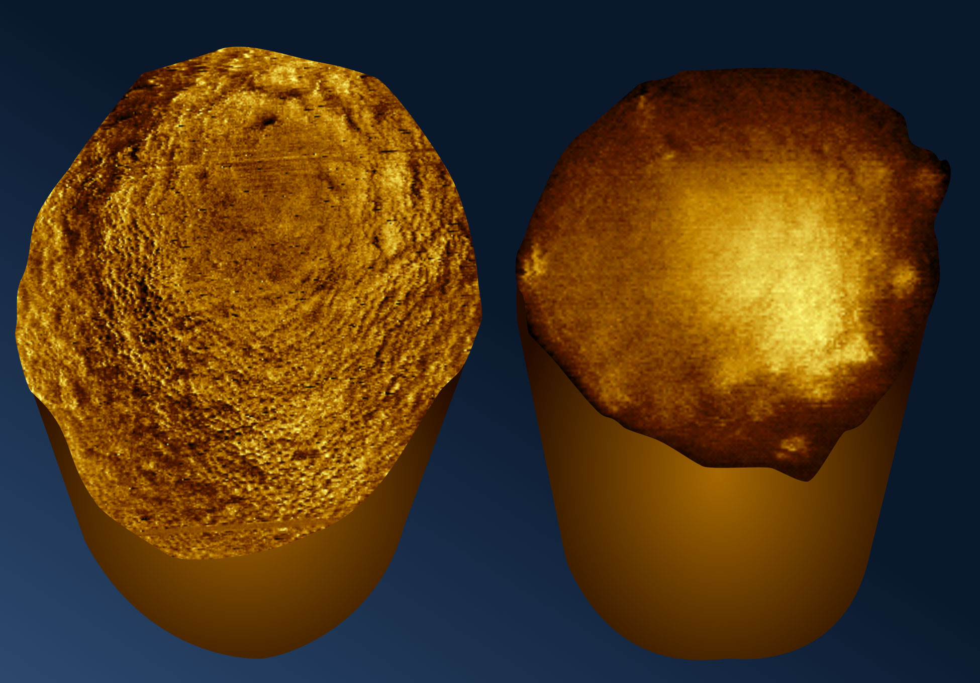
Scientists have captured the first molecular-resolution images of the ends of rod-shaped bacteria, revealing previously unseen structural features.
Scientists have captured the ends of living, rod-shaped bacteria at molecular resolution for the first time, exposing surface structures that were previously impossible to examine in such detail. The images reveal differences in bacterial architecture that could help researchers understand why some species respond to antibiotics differently from others.
The breakthrough, led by researchers at UCL (University College London) and published in Nature Communications, relied on an unexpected discovery: bacteria attached to a specially treated glass surface could remain standing upright, allowing scientists to examine their exposed tips, known as poles.
A New Way to Examine Living Bacteria
To keep bacterial cells from moving during imaging, the researchers coated glass with a chemical that binds to proteins on their surfaces. The technique held the bacteria securely without preventing them from growing or reproducing, overcoming a major obstacle to studying living cells at extremely high resolution.
After 20 minutes, the bacteria were generally immobilized horizontally. When left for longer (about 50 minutes), many unexpectedly stood upright, exposing the ends of their cells for direct examination.
The team used atomic force microscopy to image these surfaces. Unlike conventional light microscopes, this technology uses a probe just a few nanometers wide that gently taps across the cell, detecting its contours much like a finger reading Braille. The resulting measurements reveal structural details far smaller than those visible with ordinary light microscopy.
Unexpected Differences in Bacterial Surfaces
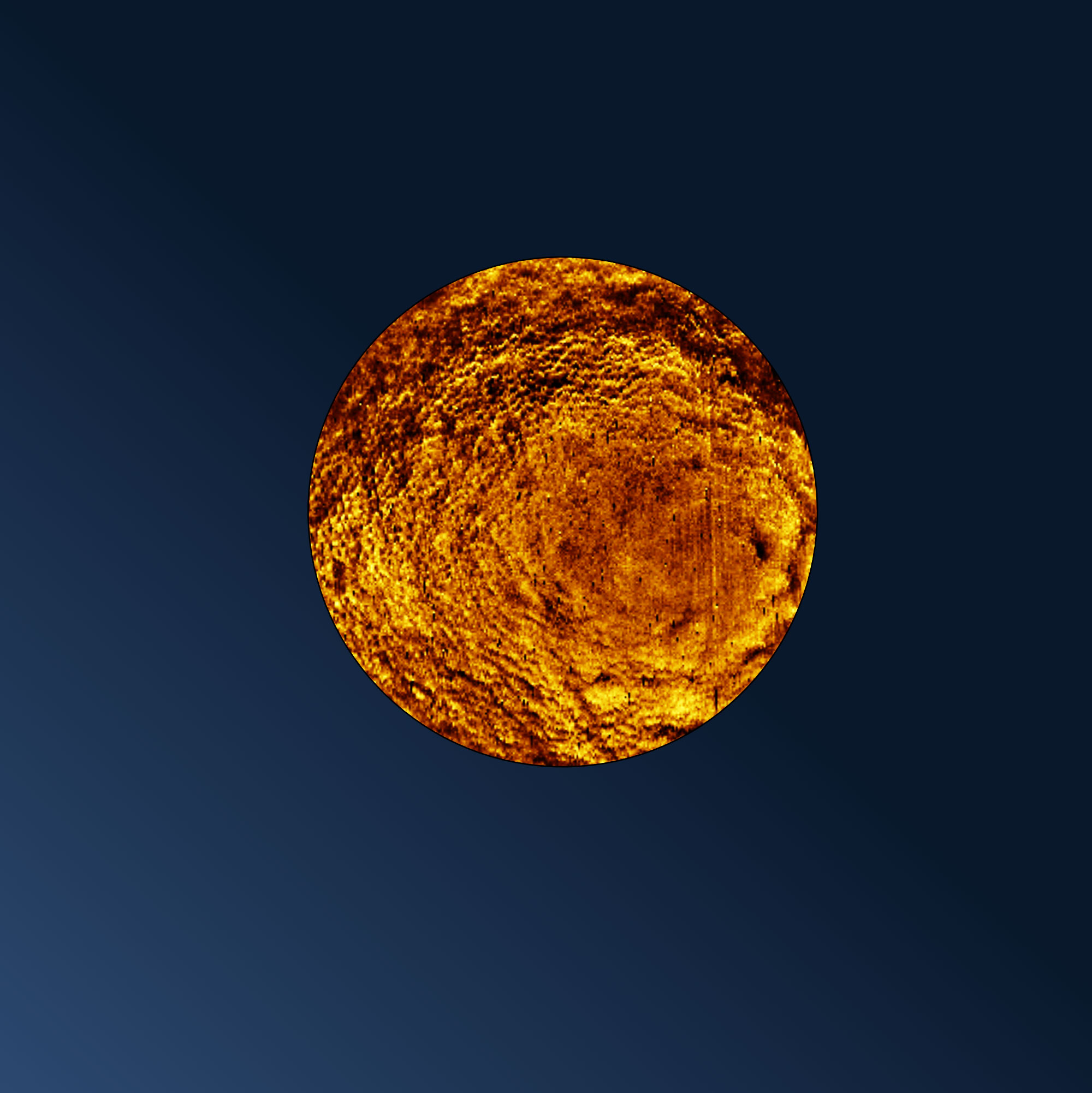
The images showed that bacterial poles change with cellular activity. Rapidly dividing bacteria (i.e., ones that were dividing rapidly) had fold-like defects at their ends, while less active cells generally displayed smoother, more rounded poles. These irregularities suggest that the bacterial surface may not have fully remodeled after cell division.
The researchers also examined E. coli (Escherichia coli) alongside Pseudomonas aeruginosa and Acinetobacter baumannii, species associated with difficult-to-treat infections. Unlike E. coli, the other two bacteria lacked the network of tiny openings (pores) observed on its surface. These structural differences may influence how the species interact with antibiotics, although their precise effects on drug susceptibility remain to be established.
Lead author Dr. Anna Scheeder, based at the London Centre for Nanotechnology at UCL, said: “Our immobilization method opens up exciting new possibilities. We can now study the armor of bacteria across different species more reliably and clearly observe nanoscale differences that were previously difficult to assess.”
Exploring a Potential Weak Point in Drug-Resistant Bacteria
Bacterial poles are closely connected to cell division, when new ends form as a bacterium splits into two daughter cells. Some antibiotics preferentially act on these regions, making their molecular structure a potential source of information for developing new treatments.
Senior author Professor Bart Hoogenboom (London Centre for Nanotechnology at UCL and UCL Physics and Astronomy) said: “Imaging the poles of bacteria is like opening a view of the back of the moon. We are looking at something that no one else has seen before. The poles are where cells divide and so contain critical information about this process. They may also be a particular target for antibiotics, with some bacteria-killing treatments preferring to attack this part of the cell.
“Our method of immobilizing the cells without affecting their ability to grow and reproduce also means we can image a much larger range of live bacteria than was previously possible. This is important as it may help us understand differences between species and why antibiotics work for some of them but not others.
“Our work is fundamental science that ultimately aims to find new leads to target drug-resistant bacteria.”
Reference: “Nanometer-resolution imaging of living bacteria across species and cellular orientations” by Anna Scheeder, Joanna Szczepaniak, Yulianna Koziy, Naomi Mara Claro De Oliveira, William Trewby, Renata Kaminska, Jeremy Brown, Colin Kleanthous and Bart W. Hoogenboom, 9 November 2026, Nature Communications.
DOI: 10.1038/s41467-026-78975-y
The work was funded by the Wellcome Trust, the Biotechnology and Biological Sciences Research Council (BBSRC), the Rosetrees Trust, and the Engineering and Physical Sciences Research Council (EPSRC).
Never miss a breakthrough: Join the SciTechDaily newsletter.
Follow us on Google and Google News.