A new study shows that truncated versions of the Tau protein are more likely to form the sticky filaments seen in the brains of people with Alzheimer’s disease.
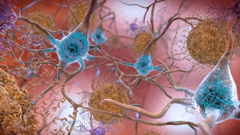
Many neurodegenerative diseases, including Alzheimer’s, are characterized by tangled proteins called Tau fibrils. In a new study, MIT chemists have gained insight into how these fibrils form, and identified a potential target for drugs that could interfere with this formation.
In the new study, the researchers discovered that one segment of the Tau protein is more flexible than expected, and this flexibility helps the fibrils take on a variety of different shapes. They also showed that these fibrils are more likely to form when the ends of the Tau protein are lopped off.
“This protein cleavage happens relatively early in Alzheimer’s disease, and that helps to speed up aggregation, which is undesirable,” says Mei Hong, an MIT professor of chemistry and the senior author of the new study.
The researchers also pinpointed a sequence of amino acids that appears to help the Tau protein bend in different directions, which they believe could make a good target for drugs that would interfere with the formation of Tau tangles.
MIT postdoc Nadia El Mammeri is the lead author of the study, which was published on July 14 in the journal Science Advances. MIT postdocs Pu Duan and Aurelio Dregni are also authors of the paper.
Fibril Formation
In the healthy brain, Tau proteins bind to microtubules and help to stabilize them. The protein contains four repeating subunits, each slightly different, known as R1, R2, R3, and R4. In the brains of people with Alzheimer’s and other neurodegenerative diseases, abnormal versions of Tau form stringy filaments that clump together, causing tangles in the brain.
Learning more about the structures of those filaments could help researchers figure out how abnormal Tau proteins become misfolded, but studying those filaments has been difficult because of their inherently disordered structure. In this study, the researchers used nuclear magnetic resonance (NMR) to determine some of those structures, using a version of the Tau protein generated in the lab using recombinant DNA.
The researchers focused on the central core of the Tau protein, where folded protein strands called beta sheets create a very rigid structure. This core is bookended by floppy segments. While the exact structure of these floppy segments is unknown, researchers have used electron microscopy to show that they form a “fuzzy coat” that surrounds the central core.
To explore what happens when those end segments are lost, as often happens in Alzheimer’s disease, the researchers chopped them off and then used NMR to analyze the resulting protein structure. Without those floppy segments, the researchers found that the rigid cores formed filaments much more easily. This suggests that the fuzzy coat helps to prevent the protein from forming filaments, which could have a protective effect against neurodegenerative disease.
“What that tells you is that fuzzy coat in the natural protein actually has a protective role. It slows down fibril formation. Once you strip away these sections, then the aggregation process happens much faster,” Hong says.
Protein Flexibility
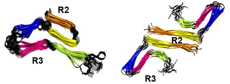
The researchers also found that the R3 repeat, which makes up much of the rigid core, is itself very rigid. However, the R2 repeat, which makes up the rest of the core, is more flexible and can produce different conformations, depending on environmental conditions such as temperature.
“This finding highlights how the environment influences the form and shape of the aggregate at the atomic level, similar to how a chameleon adapts its color to the environment. Small changes in temperature are sufficient to change the overall shape of the aggregate, which must be regarded as amazing and usually not observed in functional systems,” says Roland Riek, a professor of chemistry and applied biosciences at ETH Zurich, who was not involved in the study.
Under different conditions, R2 can exist as either a straight or hinged segment, the researchers showed. They believe this conformational flexibility may account for the slight differences in structure that have been seen in Tau proteins found in different diseases, including Alzheimer’s, corticobasal degeneration, and argyrophilic grain disease.
Within the R2 repeat, the researchers also identified a sequence of six amino acids that appear to make the structure more flexible than other R segments. This region could offer an accessible target for drugs that would inhibit the formation of Tau fibrils, Hong says.
“This region of R2 is conformationally plastic, so maybe this is a vulnerable spot that could be targeted by small molecule drugs,” she says. “The R3 region is so stable and rigid that it’s probably very hard to disaggregate Tau fibrils by focusing on that part.”
The researchers now plan to explore whether they can generate Tau structures that more closely match the structures of Tau proteins taken from the brains of patients with Alzheimer’s and other neurodegenerative diseases, by truncating the protein in specific locations or adding chemical modifications that have been linked with those diseases.
Reference: “Amyloid fibril structures of tau: Conformational plasticity of the second microtubule-binding repeat” by Nadia El Mammeri, Pu Duan, Aurelio J. Dregni and Mei Hong, 14 July 2023, Science Advances.
DOI: 10.1126/sciadv.adh4731
The research was funded by the National Institutes of Health and an NIH Ruth L. Kirschstein Individual National Research Service Award.
Never miss a breakthrough: Join the SciTechDaily newsletter.
Follow us on Google and Google News.