Using new detectors and algorithms, researchers at Cornell can now image individual atoms in 3D with record-setting resolution, limited only by the atoms’ own vibrations.
In 2018, Cornell researchers built a high-powered detector that, in combination with an algorithm-driven process called ptychography, set a world record by tripling the resolution of a state-of-the-art electron microscope.
As successful as it was, that approach had a weakness. It only worked with ultrathin samples that were a few atoms thick. Anything thicker would cause the electrons to scatter in ways that could not be disentangled.
Now a team, again led by David Muller, the Samuel B. Eckert Professor of Engineering, has bested its own record by a factor of two with an electron microscope pixel array detector (EMPAD) that incorporates even more sophisticated 3D reconstruction algorithms.
The resolution is so fine-tuned, the only blurring that remains is the thermal jiggling of the atoms themselves.
The group’s paper, “Electron Ptychography Achieves Atomic-Resolution Limits Set by Lattice Vibrations,” published May 20 in Science. The paper’s lead author is postdoctoral researcher Zhen Chen.
“This doesn’t just set a new record,” Muller said. “It’s reached a regime which is effectively going to be an ultimate limit for resolution. We basically can now figure out where the atoms are in a very easy way. This opens up a whole lot of new measurement possibilities of things we’ve wanted to do for a very long time. It also solves a long-standing problem – undoing the multiple scattering of the beam in the sample, which Hans Bethe laid out in 1928 – that has blocked us from doing this in the past.”
Ptychography Reveals Hidden Atomic Structure
Ptychography works by scanning overlapping scattering patterns from a material sample and looking for changes in the overlapping region.
“We’re chasing speckle patterns that look a lot like those laser-pointer patterns that cats are equally fascinated by,” Muller said. “By seeing how the pattern changes, we are able to compute the shape of the object that caused the pattern.”
The detector is slightly defocused, blurring the beam, in order to capture the widest range of data possible. This data is then reconstructed via complex algorithms, resulting in an ultraprecise image with picometer (one-trillionth of a meter) precision.
“With these new algorithms, we’re now able to correct for all the blurring of our microscope to the point that the largest blurring factor we have left is the fact that the atoms themselves are wobbling, because that’s what happens to atoms at finite temperature,” Muller said. “When we talk about temperature, what we’re actually measuring is the average speed of how much the atoms are jiggling.”
The researchers could possibly top their record again by using a material that consists of heavier atoms, which wobble less, or by cooling down the sample. But even at zero temperature, atoms still have quantum fluctuations, so the improvement would not be very large.
Applications: From Quantum Materials to Catalysts
This latest form of electron ptychography will enable scientists to locate individual atoms in all three dimensions when they might be otherwise hidden using other imaging methods. Researchers will also be able to find impurity atoms in unusual configurations and image them and their vibrations, one at a time. This could be particularly helpful in imaging semiconductors, catalysts and quantum materials – including those used in quantum computing – as well as for analyzing atoms at the boundaries where materials are joined together.
The imaging method could also be applied to thick biological cells or tissues, or even the synapse connections in the brain – what Muller refers to as “connectomics on demand.”
While the method is time-consuming and computationally demanding, it could be made more efficient with more powerful computers in conjunction with machine learning and faster detectors.
“We want to apply this to everything we do,” said Muller, who co-directs the Kavli Institute at Cornell for Nanoscale Science and co-chairs the Nanoscale Science and Microsystems Engineering (NEXT Nano) Task Force, part of Cornell’s Radical Collaboration initiative. “Until now, we’ve all been wearing really bad glasses. And now we actually have a really good pair. Why wouldn’t you want to take off the old glasses, put on the new ones, and use them all the time?”
Reference: “Electron ptychography achieves atomic-resolution limits set by lattice vibrations” by Zhen Chen, Yi Jiang, Yu-Tsun Shao, Megan E. Holtz, Michal Odstrcil, Manuel Guizar-Sicairos, Isabelle Hanke, Steffen Ganschow, Darrell G. Schlom and David A. Mull, 21 May 2021, Science.
DOI: 10.1126/science.abg2533
Co-authors include Darrell Schlom, the Herbert Fisk Johnson Professor of Industrial Chemistry; Yi Jiang, Ph.D. ’18 and now a beamline data scientist at Argonne National Laboratory; postdoctoral researchers Yu-Tsun Shao and Megan Holtz, Ph.D. ’17; and researchers from the Paul Scherrer Institute and the Leibniz Institute for Crystal Growth.
The research was supported by the National Science Foundation through Cornell’s Platform for the Accelerated Realization, Analysis and Discovery of Interface Materials (PARADIM). The researchers also made use of the Cornell Center for Materials Research, which is supported by the NSF’s Materials Research Science and Engineering Center program.
Never miss a breakthrough: Join the SciTechDaily newsletter.
Follow us on Google and Google News.
21 Comments
This is a biggie across many fields.
So is this big news or ultra small news?
Big news about small stuff!
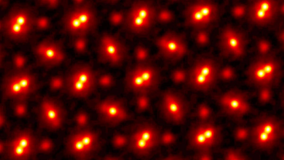
I would have liked it if they had explained what we’re looking at in that image. I get it that they’re atoms, but which dots correspond to which atoms? It would help me visualize what a PrSc03 crystal structure actually looks like.
What you are seeing is 2 atoms of praseodymium(Pr) bonded together(the 2 bright dot-pairs) with scandium atoms(single dull red dots) sandwiched in a lattice with the Pr pairs. What can’t really be seen are the oxygen atoms that surround the Praseodymium atom-pairs. The oxygen atoms are much smaller than the Pr and Sc atoms themselves.
Here is a link that graphically shows the lattice system. Really helpful.https://core.ac.uk/download/pdf/52920025.pdf
Garin
That is an interesting interpretation. Why are there praseodymium pairs when the chemical formula doesn’t reflect that? If the unit cell contains two Pr-pairs, why aren’t there doubled Sc as well?
Good questions!
The chemical formula is reduced, so is not representing the unit cell, c.f. diamond cubic lattice of C that has 8 atoms in the unit cell.
As far as I can interpret the crystal structure representation [thanks Garin!] you are looking at a (pseudo)cubic lattice of ABX3 where each larger Pr atom is surrounded by an octahedron of Sc atoms [ https://en.wikipedia.org/wiki/Perovskite_(structure) ]. The Pr and and Sc pairs is an artifact of the image, but we see only the front Sc of an Sc pair latticed with a clearly visible Pr pair (and the cut is done so there is a lacking Pr pair).
I would guess that there is a lot of aliasing and it looks like rotations going on in the electron ptychographic reconstruction that we compare with, either real or enforced by the method, so it is an iffy comparison.
Could they not also sub freeze the sample to slow the atom down??? Reduce the wiggles for even better image of atoms..
If this is an image of atoms how come we don’t see the atoms of what is in the space before the supposed layer of atoms that’s being recorded? Even in a vacuum there is something!
I think that would be subatomic and quantum scale particles you’re thinking of
A vacuum means there is nothing.
The large ones are responsive to the instalment.
Instrument
Wow, How small can you go? Now I know!
What about uncertainty?
Scitechdaily just a bunch of uberimmaginations running unrestrained and unsupervised. Mostly just fake garbage imaginative type bull
I’ve heard there is a supercamera that puts the speed of light in slow motion. That surely will take clearer picture of an atom wobbling. So that we would now know that atoms are actually flat. PicoKPH?
Looks like tiny evil little eyes…. I wonder if this is why my cats go nuts for what seems like no reason at all???
So is the space in between the atoms considered dark matter? I know nothing btw so this is a serious question….
I too was wondering exactly what crystal structure we were viewing. I had assumed the pairs were only one atom with a p-valence electron. Therefore concluding this was a simple salt as the circular atom could be a group one atom with an S valence. The truth is much more interesting!