
Close Menu
    Facebook X (Twitter) Instagram
    SciTechDaily
    • Biology
    • Chemistry
    • Earth
    • Health
    • Physics
    • Science
    • Space
    • Technology
    Facebook X (Twitter) Pinterest YouTube RSS
    SciTechDaily
    Home»Health»Innovative X-Ray Imaging Directly Shows COVID-19 Can Cause Vascular Damage to the Heart
    Health

    Innovative X-Ray Imaging Directly Shows COVID-19 Can Cause Vascular Damage to the Heart

    By University of GöttingenDecember 30, 20211 Comment4 Mins Read
    Facebook Twitter Pinterest Telegram LinkedIn WhatsApp Email Reddit
    Share
    Facebook Twitter LinkedIn Pinterest Telegram Email Reddit
    Heart Tissue Severe COVID-19
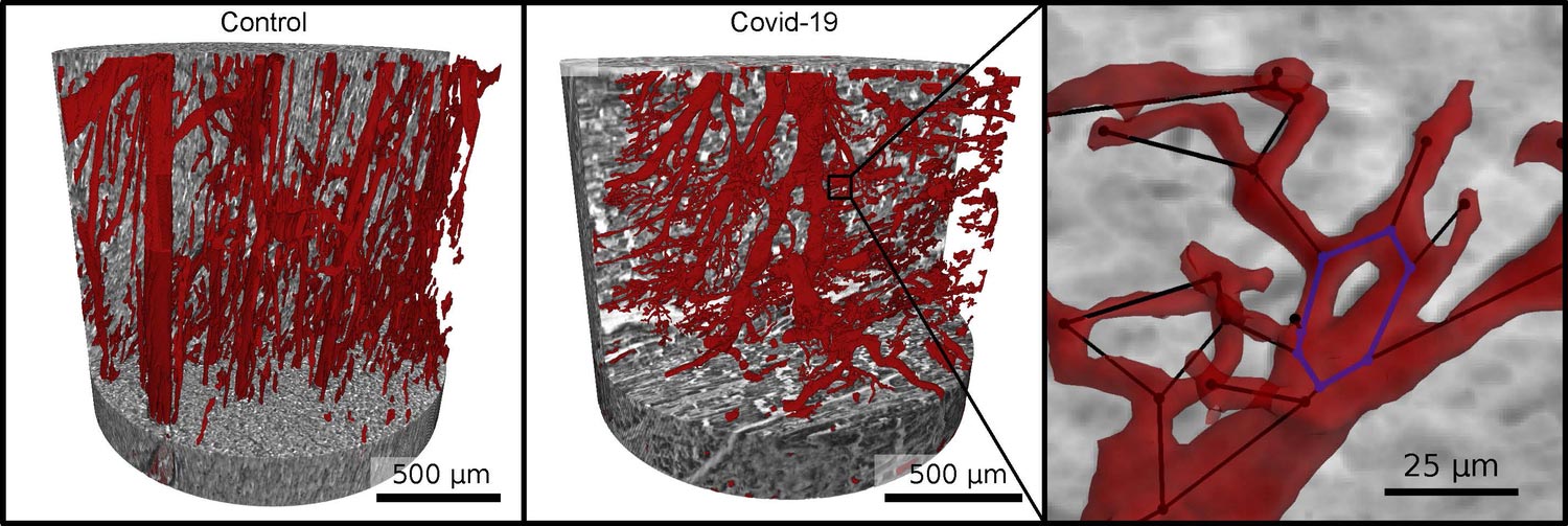
    Vascular network (red) in healthy heart tissue (left) and in severe COVID-19 (right). Due to faulty reforming of the network as a result of COVID-19, numerous branches, splits and even loops develop in the capillaries, which can be analyzed mathematically. Credit: M. Reichardt, P. Møller Jensen, T. Salditt

    Interdisciplinary research team from Göttingen University and Hannover Medical School are first to prove this directly.

    An interdisciplinary research team from the University of Göttingen and Hannover Medical School (MHH) has detected significant changes in the heart muscle tissue of people who died from COVID-19. Damage to lung tissue has been the research focus in this area for some time and has now been thoroughly investigated. The current study underpins the involvement of the heart in COVID-19 at the microscopic level for the first time by imaging and analyzing the affected tissue in the three dimensions. The results were published in the journal eLife.

    The scientists imaged the tissue architecture to a high resolution using synchrotron radiation – a particularly bright X-ray radiation – and displayed it three-dimensionally. To do this, they used a special X-ray microscope that the University of Göttingen set up and operates at the German Electron Synchrotron DESY in Hamburg. They observed clear changes at the level of the capillaries (the tiny blood vessels) in the heart muscle tissue when they examined the effects there of the severe form of COVID-19 disease.
    Heart Tissue COVID-19
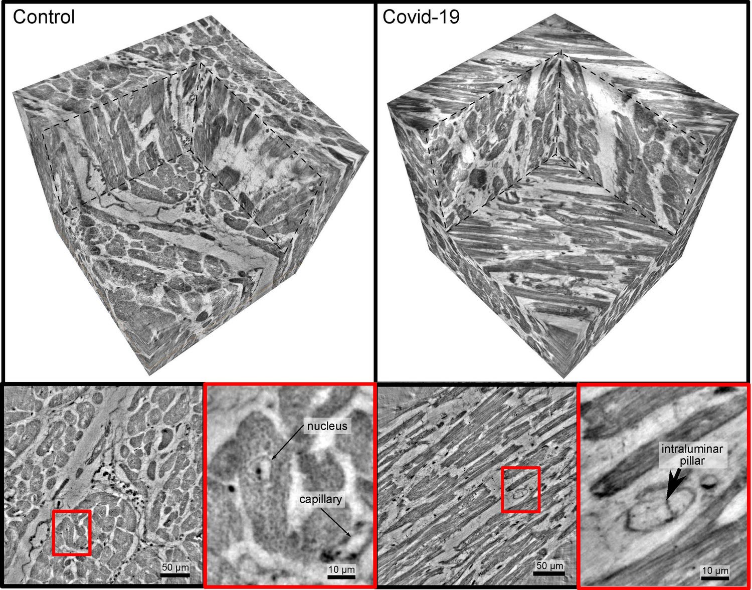
    Healthy heart tissue (left) and in severe COVID-19 (right). While the blood vessels in the healthy case show an intact structure (bottom right), one can see cavities or pouches and tiny tubular structures forming in the fine blood vessels (capillaries) in the COVID-19 tissue sample, which indicate particular morphological changes. Credit: M Reichardt, T Salditt

    In comparison with a healthy heart, X-ray imaging of tissues affected by severe disease, revealed a network full of splits, branches, and loops which had been chaotically remodeled by the formation and splitting of new vessels. These changes are the first direct visual evidence of one of the main drivers of lung damage in COVID-19: a special kind of “intussusceptive angiogenes” (meaning new vessel formation) in the tissue.

    In order to visualize the capillary network, the vessels in the three-dimensional volume first had to be identified using machine learning methods. This initially required researchers to painstakingly, manually label the image data. “To speed up image processing, we therefore also automatically broke the tissue architecture down into its local symmetrical features and then compared them,” explains Marius Reichardt, at the University of Göttingen and first author of the paper. “The parameters obtained from this then showed a completely different quality compared to healthy tissue, or even to diseases such as severe influenza or common myocarditis,” explain the leaders of the study, Professor Tim Salditt from the University of Göttingen and Professor Danny Jonigk from the MHH.

    There is a very special feature of this study: in contrast to the vascular architecture, the required data quality could be achieved using a small X-ray source in the laboratory of the University of Göttingen. In principle, this means it could also be done in any clinic to support pathologists with routine diagnostics. In the future, the researchers want to further expand the approach of converting the characteristic tissue patterns into abstract mathematical values in order to develop automated tools for diagnostics, again by further developing laboratory X-ray imaging and validating it with data from synchrotron radiation. The collaboration with DESY will be further expanded in the coming years.

    Reference: “3D virtual histopathology of cardiac tissue from Covid-19 patients based on phase-contrast X-ray tomography” by Marius Reichardt, Patrick Moller Jensen, Vedrana Andersen Dahl, Anders Bjorholm Dahl, Maximilian Ackermann, Harshit Shah, Florian Länger, Christopher Werlein, Mark P Kuehnel, Danny Jonigk and Tim Salditt, 21 December 2021, eLife.
    DOI: 10.7554/eLife.71359

    Never miss a breakthrough: Join the SciTechDaily newsletter.
    Follow us on Google and Google News.

    Cardiology COVID-19 Heart Infectious Diseases University of Göttingen
    Share. Facebook Twitter Pinterest LinkedIn Email Reddit

    Related Articles

    Small Study Finds COVID-19 Vaccine Safe in Patients With Previous Myocarditis

    People With Heart Defects at Greater Risk for Severe COVID-19 Illness and Death

    COVID Spike Protein Binds to Heart’s Vascular Cells – May Contribute to Severe Microvascular Damage

    COVID-19 Infections Increase Risk of Serious Heart Conditions Up to a Year Later

    Heart Damage After COVID-19 Uncommon in College Athletes

    Low-Dose Aspirin Use for Heart Disease May Reduce Likelihood of COVID-19 Infection

    Research Reveals How COVID-19 Can Kill Heart Muscle Cells, Interfere With Contraction

    Heart Damage Found in More Than Half of COVID-19 Patients Discharged From Hospital

    Cardiac Ultrasounds Show Damaging Impact of COVID-19 on the Heart

    1 Comment

    1. Claire on December 30, 2021 12:05 pm

      Dr. Vladimir Zelenko exposed executives that are taking hydroxychloroquine and ivermectin prophylactically. They are not taking the vaccine! These drugs are all on the World Health Organization’s “Model List of Essential Medicines” website. Unfortunately doctors are shamed, black mailed by certain individuals and even SUSPENDED by administration in order not to prescribe these medicines. Get your Ivermectin today while you still can https://medicalfreedom.wiki

      Reply
    Leave A Reply Cancel Reply

    • Facebook
    • Twitter
    • Pinterest
    • YouTube

    Don't Miss a Discovery

    Subscribe for the Latest in Science & Tech!

    Trending News

    Could Low Vitamin D Be Making Your Pain Worse?

    Scientists Discover Once-Weekly Workout That Melts Belly Fat Surprisingly Effectively

    Scientists Just Tested a Thruster Powerful Enough for Human Missions to Mars

    Doctors Say Your Ice Pack Might Be Making Injuries Worse

    Scientists Discover 43-Foot Sea Reptile Twice the Size of a Great White Shark

    Bees and Birds Are Drinking Alcohol From Flowers

    Scientists Discover How Obesity May Trigger Alzheimer’s Disease

    Scientists Confirm Alcohol Causes Widespread Health Damage

    Follow SciTechDaily
    • Facebook
    • Twitter
    • YouTube
    • Pinterest
    • Newsletter
    • RSS
    SciTech News
    • Biology News
    • Chemistry News
    • Earth News
    • Health News
    • Physics News
    • Science News
    • Space News
    • Technology News
    Recent Posts
    • Just Minutes of Intense Exercise Could Slash Disease Risk by 50%
    • People Who Stop Ozempic Often Don’t Gain the Weight Back
    • Why More People in Their 30s Are Suddenly Getting Colon Cancer
    • Scientists Discover Sperm Seem To Bypass a Fundamental Law of Physics
    • The Secret Behind Indigenous Andeans’ “Digestive Superpower” May Be Potatoes
    Copyright © 1998 - 2026 SciTechDaily. All Rights Reserved.
    • Science News
    • About
    • Contact
    • Editorial Board
    • Privacy Policy
    • Terms of Use

    Type above and press Enter to search. Press Esc to cancel.