Using an FDA-approved contrasting agent containing tiny gas-filled microbubbles, coupled with a noninvasive ultrasound device that targets precise locations of the brain, a team of neurobiologists showed that repeated treatments temporarily opened the blood-brain barrier while causing no structural or functional damage.
Researchers used a noninvasive, focused ultrasound device to temporarily disrupt the blood-brain barrier and target discrete regions of the brain. This approach may open the way for new drug delivery strategies for diseases such as brain cancers and Alzheimer’s, conditions typically not amenable to more conventional treatments.
Using an FDA-approved contrasting agent containing tiny gas-filled microbubbles, coupled with an ultrasound platform that targeted precise locations in the brains of nonhuman primates, researchers from Harvard Medical School showed that repeated treatments temporarily opened the blood-brain barrier while causing no structural or functional damage. Their results, which were published online in the journal Cancer Research, support clinical testing of this new application.
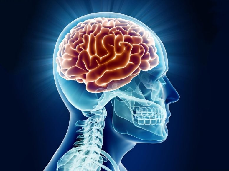
The brain is protected from most toxins and bacterial infections by the blood-brain barrier (BBB), a physical separation between the brain and the circulatory system which normally only permits small, essential molecules like oxygen and glucose through. Large molecules like chemotherapy agents and antibiotics cannot easily permeate the BBB, making it one of the primary obstacles to treating diseases of the brain.
To address this problem, Nathan McDannold, HMS associate professor of radiology at the Brigham and Women’s Hospital and first author on the paper, and Margaret Livingstone, HMS professor of neurobiology, introduced a microbubble contrast agent into the circulatory system of nonhuman primates. Guided by an MRI, they then targeted specific areas of the brain with a device comprised of an array of 1,024 directed ultrasound transmitters, using short bursts at low power. Due most likely to the mechanical stimulation of microbubbles by ultrasound frequencies, the BBB weakened at targeted areas enough for the normally impermeable imaging dye to penetrate the brain. This effect lasts for roughly four hours, and is disruptive enough for drugs and large molecules to enter the brain and target, say, tumors.
“This may really change the way that people use drugs in the brain,” said McDannold. “We have this treatment that is totally non-invasive, and you can repeat it over and over again.”
The researchers also verified the safety of this method. After multiple sessions of repeated disruptions of the BBB in the visual cortex and the central visual field, the animals performed a series of visual discrimination tasks that included learning and recognizing different symbols, at varying sizes, for a reward.
“They didn’t have any trouble with memory. They were smart, and were just as fast and adept at it as they were before. They could discriminate 26 different tiny symbols,” said Livingstone, who ran the tests. “We couldn’t pick up anything wrong.”
The anatomical features of the targeted areas in the brain also showed no damage or inflammation, and the only evidence of treatments were a few injured capillaries and small groups of leaked blood cells. “My hope is that the time window we disrupt it for is long enough for drugs to get in but short enough not to cause damage,” said McDannold. “But compared to radiation or injecting drugs, even catheterization, it’s a risk worth taking.”
While further tests are needed to completely eliminate potential hazards, repeated treatments with the current system have shown no evidence of injury.
The team plans on improving the technology, particularly in targeting guidance and cost reduction. Because the focused ultrasound device they used is already FDA approved for other uses, and safety has been demonstrated, they are optimistic that this technique is ready for clinical trials. “We’re ready to move this to patients,” said McDannold. “This data that we’ve gotten over the past couple of years is really convincing to us.”
Reference: ” Temporary Disruption of the Blood–Brain Barrier by Use of Ultrasound and Microbubbles: Safety and Efficacy Evaluation in Rhesus Macaques” by Nathan McDannold, Costas D. Arvanitis, Natalia Vykhodtseva and Margaret S. Livingstone, 15 July 2012, Cancer Research.
DOI: 10.1158/0008-5472.CAN-12-0128
This work was supported by award number RC2NS069413 from the National Institute of Neurological Disorders And Stroke.
Never miss a breakthrough: Join the SciTechDaily newsletter.
Follow us on Google and Google News.