Unlocking Cancer Drug Targets Through Protein Complex Analysis
Three proteins work together to transmit signals for cell division, revealing new targets for cancer-fighting drugs.
Cell replication in our bodies is triggered by a cascade of molecular signals transmitted between proteins. Compounds that block these signals when they run amok show potential as cancer drugs. Recently, scientists uncovered the molecular mechanisms that underlie a step in the signal transmission pathway that requires three proteins to link up.
The detailed knowledge about this three-protein complex, determined with synchrotron X-ray user facilities, points the way to new targets for drugs that fight certain types of cancer.
Advancements in Understanding Cancer Signaling Pathways
Some promising anticancer drugs work by jamming proteins that transmit signals for cells in the body to replicate. This slows the growth of tumors. However, drug-resistance mechanisms enable the signals to bypass the jam. Scientists working on cancer treatments therefore need to gain a molecular-level understanding of the ways signaling proteins interact with each other.
In this study, scientists used biochemical experiments combined with protein-structure studies to understand the details of a key step in the signaling pathway. The results provide a sharper picture of a process that had remained unclear despite decades of study. This could lead to improved drugs for lung, colorectal, pancreatic, and other cancers.
In-Depth Analysis of a Key Protein Complex
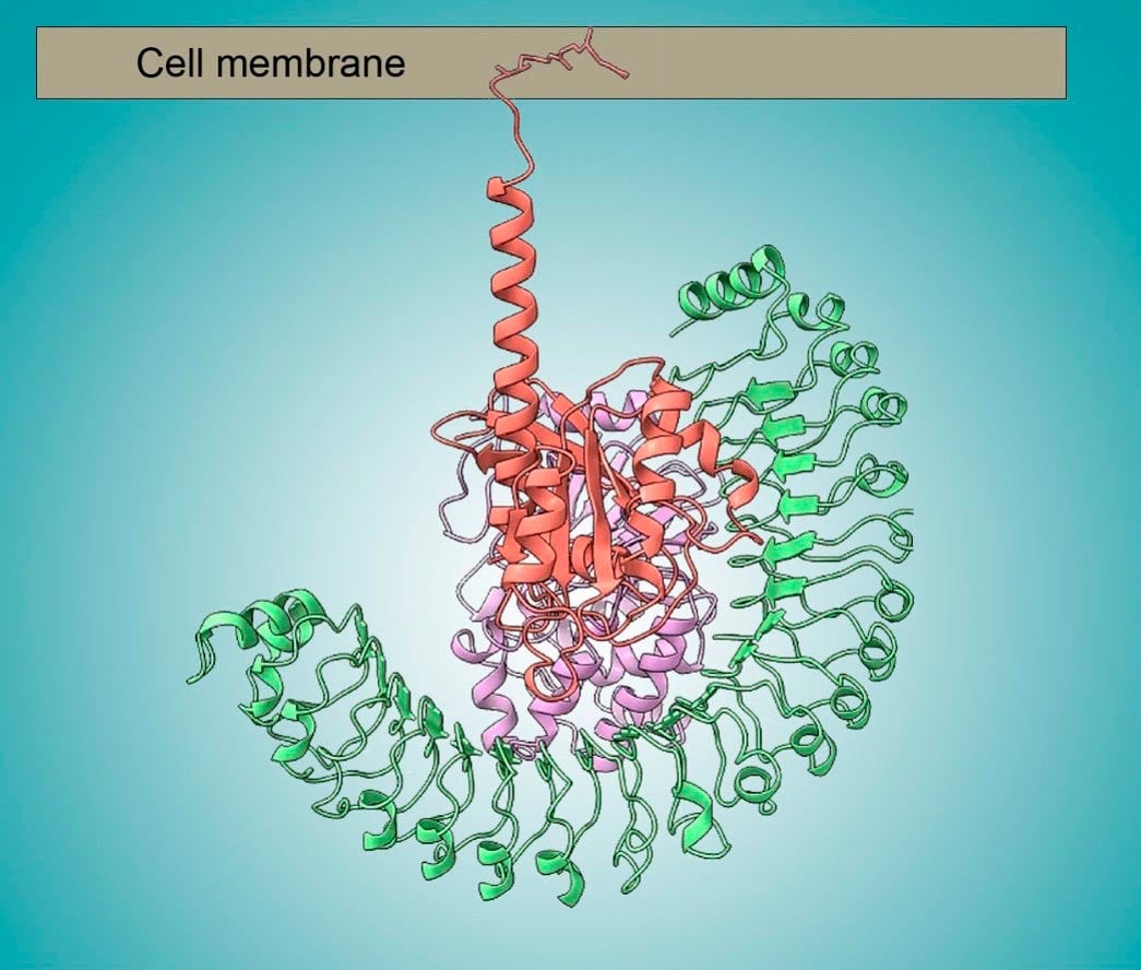
This work focused on one link in the cell-replication signaling chain, involving proteins known as SHOC2, PPIC, and RAS. When assembled, this three-protein complex becomes chemically active, enabling the next step in the signal cascade.
To obtain detailed information about where individual atoms in the proteins are located, the research team used electron microscopy at Genentech and protein crystallography at the Stanford Synchrotron Radiation Laboratory. To understand how the three proteins fit together like a jigsaw puzzle, the researchers used a technique called small-angle X-ray scattering (SAXS) at the Advanced Light Source, a Department of Energy Office of Science user facility at Lawrence Berkeley National Laboratory.
Using the SAXS data, the researchers were able to capture snapshots of the large, flexible protein complex in its native form (suspended in solution). This allowed them to model the flexibility of SHOC2, which acts as a scaffold for the other two proteins.
Implications for Future Cancer Therapies
Together with the other structural data, biochemical studies, and computer simulations, the work answered many outstanding questions, including how disease-relevant mutations affect assembly of the complex and how the proteins collectively work to activate the next step in the signaling process. In general, the work establishes fresh avenues for the discovery of new classes of targeted anticancer drugs.
Reference: “Structural basis for SHOC2 modulation of RAS signalling” by Nicholas P. D. Liau, Matthew C. Johnson, Saeed Izadi, Luca Gerosa, Michal Hammel, John M. Bruning, Timothy J. Wendorff, Wilson Phung, Sarah G. Hymowitz and Jawahar Sudhamsu, 29 June 2022, Nature.
DOI: 10.1038/s41586-022-04838-3
This research was performed at the Advanced Light Source and the Stanford Synchrotron Radiation Laboratory, both of which are Department of Energy (DOE) Office of Science user facilities. Other funding sources included the DOE Office of Science, Biological and Environmental Research program, the National Institutes of Health, and Genentech.
Never miss a breakthrough: Join the SciTechDaily newsletter.
Follow us on Google and Google News.