
New imaging research challenges a long-standing assumption about Parkinson’s disease.
A clinical imaging study from Finland suggests that rest tremor in Parkinson’s disease is not caused by greater dopamine loss. Instead, tremor appears to be linked to relatively preserved dopamine function.
Researchers at the University of Turku and Turku University Hospital analyzed clinical records along with dopamine transporter (DAT) imaging from 414 patients in Finland. These individuals were evaluated in routine care for unclear cases of parkinsonism or tremor, which makes the results highly relevant to everyday clinical practice. The findings were published in Neurology, the journal of the American Academy of Neurology.
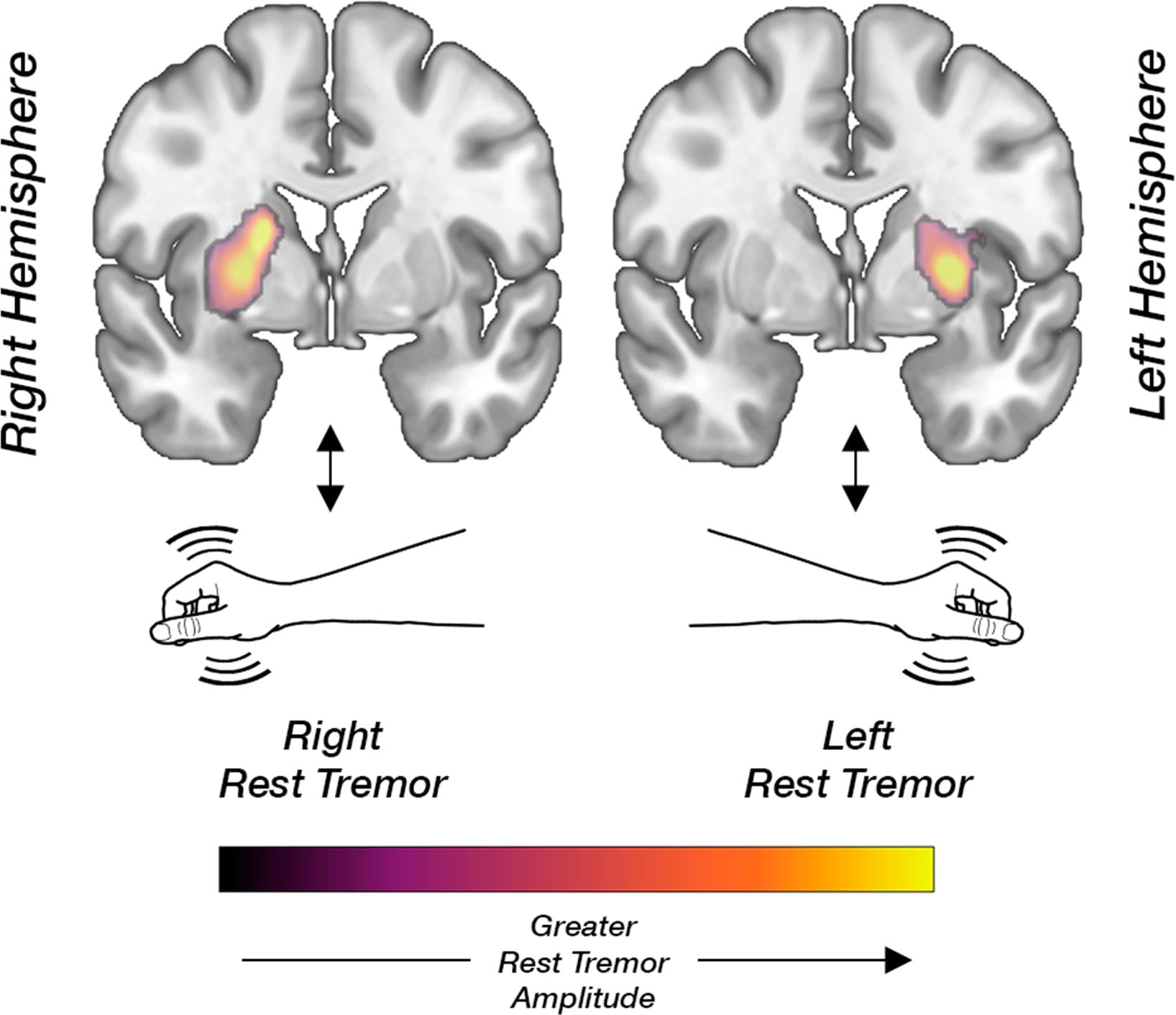
Parkinson’s disease is defined by three main motor symptoms: slowed movement (bradykinesia), muscle stiffness (rigidity), and rest tremor. Bradykinesia and rigidity are known to result from the loss of dopamine-producing neurons. Because many brain pathways cross, this damage is usually seen in the striatum on the side opposite the symptoms. However, the biological basis of rest tremor has remained unclear.
Unexpected Imaging Findings
The researchers identified a consistent pattern. Patients with rest tremor showed higher dopamine transporter binding in the striatum on the same side as the tremor. In contrast, other key motor symptoms followed the expected pattern and were linked to dopamine loss in the opposite hemisphere.
“These results show that more severe rest tremor is not simply a marker of more advanced damage to the dopamine system,” says the lead author, Neurologist Kalle Niemi, MD, PhD. “Tremor appears to involve a partly distinct neurobiological mechanism.”
The results also support earlier findings from the team based on data from the international Parkinson’s Progression Markers Initiative (PPMI). That earlier work introduced a new imaging analysis method developed by the researchers. Reproducing the same results in a separate, clinically representative group strengthens confidence in the findings.
“Our findings support the view that different symptoms of Parkinson’s disease may be driven by partly distinct neural network and neurotransmitter mechanisms,” Niemi explains. “This may help explain why tremor behaves differently from symptoms such as bradykinesia.”
Beyond Motor Symptoms
Using the same approach, the researchers also found that several non-motor symptoms of Parkinson’s disease, including depression, anxiety, and REM sleep behavior disorder, are mainly associated with monoaminergic systems other than dopamine.
Overall, the study highlights Parkinson’s disease as a complex brain disorder that involves multiple neural networks and neurotransmitter systems.
A clearer understanding of how different symptoms arise could eventually lead to more targeted and personalized treatments.
Reference: “Striatal Dopamine Transporter and Rest Tremor in Parkinson DiseaseA Clinical Validation” by Kalle J. Niemi, Elina Jaakkola, Elina Maaria Myller, Mikael R.E. Eklund, Simo Nuuttila, Tuomas Mertsalmi, Kirsi-Marja Murtomäki, Reeta Levo, Tomm Noponen, Toni Ihalainen, Filip Scheperjans, Juho Joutsa and Valtteri Kaasinen, 19 March 2026, Neurology.
DOI: 10.1212/WNL.0000000000214811
Never miss a breakthrough: Join the SciTechDaily newsletter.
Follow us on Google and Google News.