A newly identified interaction between ovarian cancer cells and surrounding abdominal cells helps explain the cancer’s unusually fast and aggressive spread.
Ovarian cancer is the deadliest gynecologic cancer, largely because it is so often discovered late. Early symptoms can be subtle and easily mistaken for common digestive or hormonal changes, so many people are diagnosed only after the disease has already spread across the abdomen.
A study from Nagoya University, published in Science Advances, points to a key reason the cancer can move so quickly once it reaches that stage. The researchers found that ovarian cancer cells can recruit mesothelial cells, the thin protective cells that normally coat the inside of the abdominal cavity and help organs glide smoothly as the body moves. Instead of remaining a passive lining, these mesothelial cells become active partners in invasion.
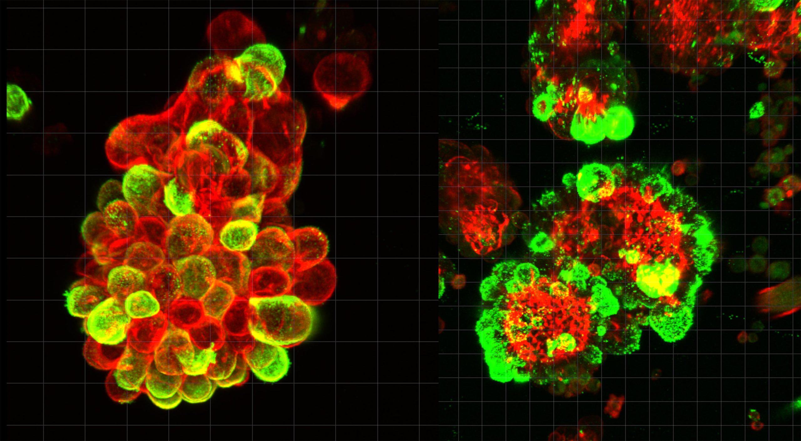
When the team examined abdominal fluid from patients, they saw that cancer cells often travel in company. Rather than floating as single cells, they frequently latch onto mesothelial cells and form mixed spheres. In the patient samples, about 60% of these cancer spheres contained recruited mesothelial cells. The cancer cells release a signaling protein called TGF-β1, which alters the mesothelial cells and prompts them to grow sharp, tissue cutting protrusions.
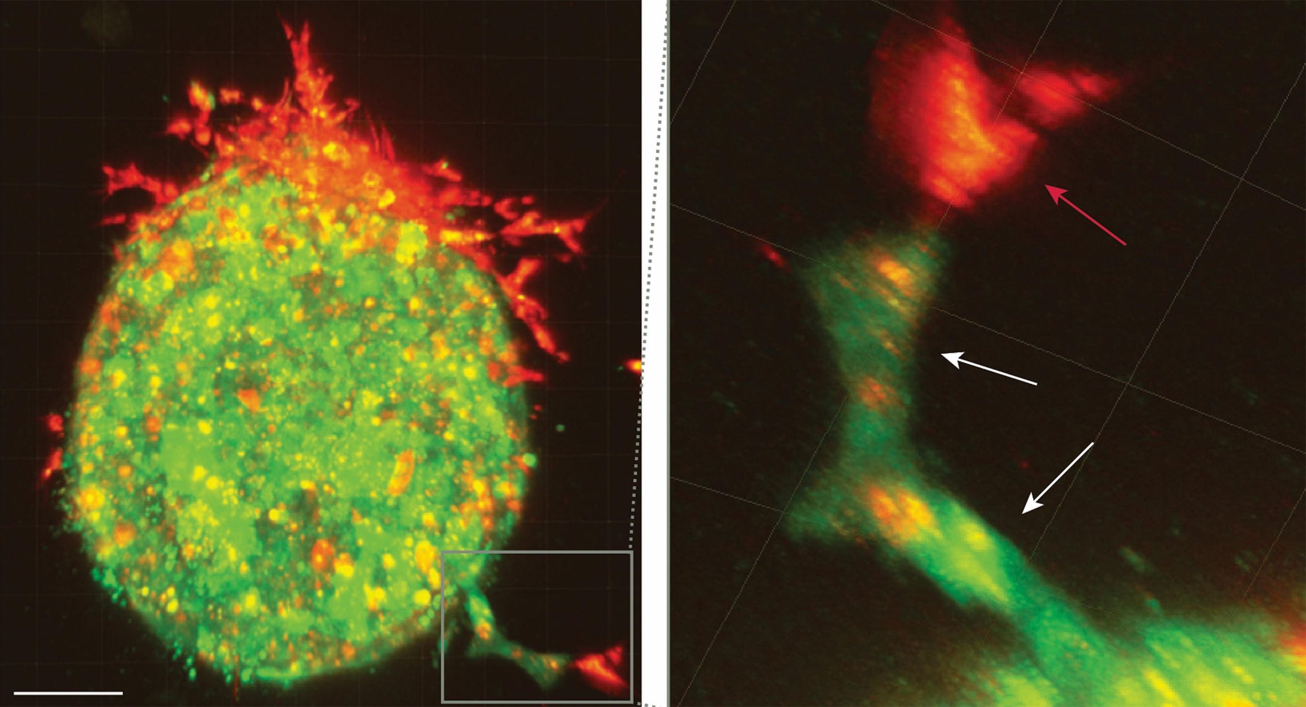
Invadopodia, spike structures that do the digging for cancer
Invadopodia, spike-like structures, do the physical drilling. In these mixed clusters, mesothelial cells do much of the digging into nearby tissue, and cancer cells slip through the openings and pathways they create. Together, the cluster invades faster and resists chemotherapy better than cancer cells alone.
This strategy matches the unusual way ovarian cancer spreads. Cells can break off from a tumor and enter abdominal fluid, where breathing, posture, and everyday movement carry them across surfaces until they find a place to attach—without needing access to the bloodstream.
That differs from cancers such as breast or lung cancer, which often spread by entering blood vessels and traveling through circulation to distant organs. Because blood follows defined routes, doctors can sometimes use blood tests to help monitor those cancers. Ovarian cancer, by contrast, moves through an internal fluid environment with no fixed path. This work suggests the cancer may exploit the abdomen’s protective lining by partnering with shed mesothelial cells during this floating stage.
During that period, before cells attach to new organs, the team found that ovarian cancer cells recruit mesothelial cells that have sloughed off the abdominal lining. The two cell types adhere to form hybrid spheres. Mesothelial cells extend invadopodia to penetrate tissue, helping the spheres invade more quickly and withstand chemotherapy once they land on organ surfaces.
Outsourcing the hard work of cell invasion
The researchers examined abdominal fluid from ovarian cancer patients using advanced microscopy to watch this process in real time. They confirmed their findings with mouse models and single-cell genetic analysis.
Lead author Dr. Kaname Uno, a former PhD student and current Visiting Researcher at Nagoya University’s Graduate School of Medicine, explained that the cancer cells do not need to become more invasive themselves. “They manipulate mesothelial cells to do the tissue invasion work. They undergo minimal genetic and molecular changes and just migrate through the openings that mesothelial cells create.”
Dr. Uno worked as a gynecologist for eight years before he pursued research. One of his patients changed his career path. She had clear screening results just three months before doctors found advanced ovarian cancer. Current medical tools failed to detect the cancer early enough to save her life. This motivated Dr. Uno to investigate why ovarian cancer spreads so rapidly.
This discovery opens new treatment possibilities. Current chemotherapy targets cancer cells but ignores the mesothelial accomplices. Future drugs could block the TGF-β1 signal or prevent the formation of these dangerous partnerships. The research also suggests that doctors could monitor these cell clusters in abdominal fluid to predict disease progression and treatment response.
Reference: “Mesothelial cells promote peritoneal invasion and metastasis of ascites-derived ovarian cancer cells through spheroid formation” by Kaname Uno, Masato Yoshihara, Yoshihiko Yamakita, Kazuhisa Kitami, Shohei Iyoshi, Mai Sugiyama, Yoshihiro Koya, Tomihiro Kanayama, Haruhito Sahara, Satoshi Nomura, Kazumasa Mogi, Emiri Miyamoto, Hiroki Fujimoto, Kosuke Yoshida, Satoshi Tamauchi, Akira Yokoi, Nobuhisa Yoshikawa, Kaoru Niimi, Yukihiro Shiraki, Jonas Sjölund, Hidenori Oguchi, Kristian Pietras, Atsushi Enomoto, Akihiro Nawa, Hiroyuki Tomita and Hiroaki Kajiyama, 6 February 2026, Science Advances.
DOI: 10.1126/sciadv.adu5944
Never miss a breakthrough: Join the SciTechDaily newsletter.
Follow us on Google and Google News.