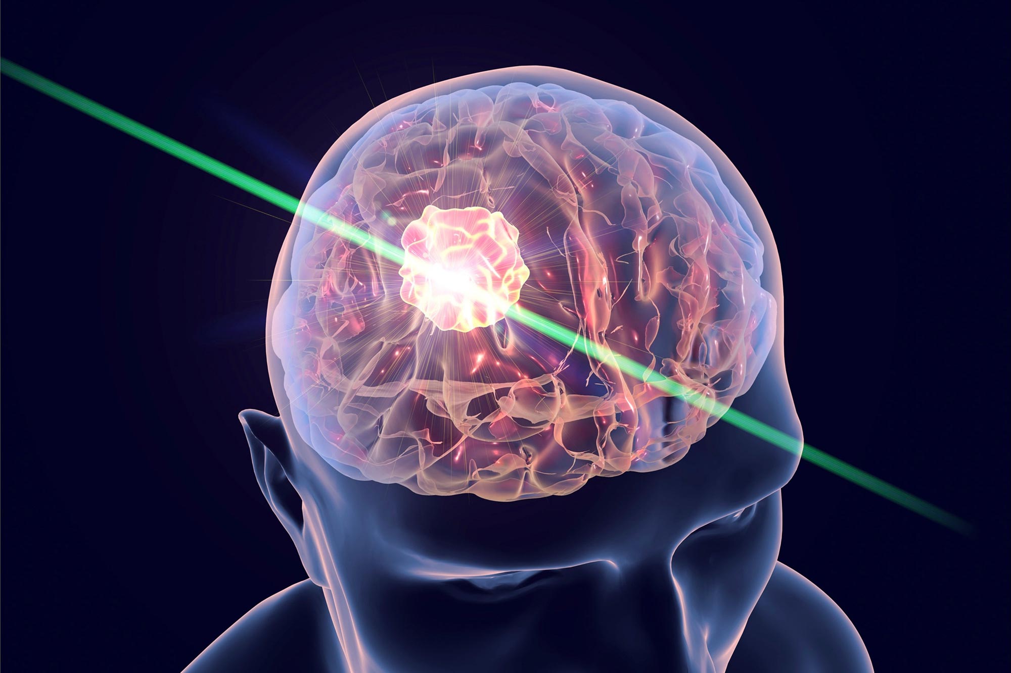
A new “molecular flashlight” technique allows non-invasive insight into brain pathologies, lighting up the future of neurological research.
- The probe can reach deep into the brain with minimal damage, earning its designation as a minimally invasive device. It emits an ultra-thin beam of light.
- By illuminating nerve tissue, the light provides detailed information about its chemical composition, enabling the detection of molecular changes caused by tumors or other lesions.
- Known as a “molecular flashlight,” this tool is currently used in research settings, but scientists hope it will eventually be applied to patient care.
- The findings are detailed in the journal Nature Methods.
Revolutionary “Molecular Flashlight” Technology
Studying molecular changes in the brain caused by cancer and neurological disorders without invasive procedures has long been a challenge in biomedical research. Now, scientists have developed a groundbreaking technique that uses an ultra-thin probe to introduce light into the brains of mice, enabling detailed molecular analysis. The findings, published today (December 31) in the journal Nature Methods, are the result of collaboration between international researchers, including teams from the Spanish National Cancer Research Centre (CNIO) and the Spanish National Research Council (CSIC).
The researchers call this innovation a “molecular flashlight” because it illuminates nerve tissue, revealing its chemical composition. This approach allows scientists to detect molecular changes associated with brain tumors—both primary and metastatic—as well as injuries like traumatic brain damage.
Minimal Invasion, Maximum Insight
The molecular flashlight is a probe less than 1 mm thick, with a tip just one micron wide—about one-thousandth of a millimeter—and invisible to the naked eye. It can be inserted deep into the brain without causing damage (for comparison, a human hair measures between 30 and 50 microns in diameter).
This flashlight-probe is not yet ready to be tested in patients, and for now, it is primarily a “promising” research tool in animal models that allows “monitoring molecular changes caused by traumatic brain injury, as well as detecting diagnostic markers of brain metastasis with high accuracy,” explain the authors of the paper.
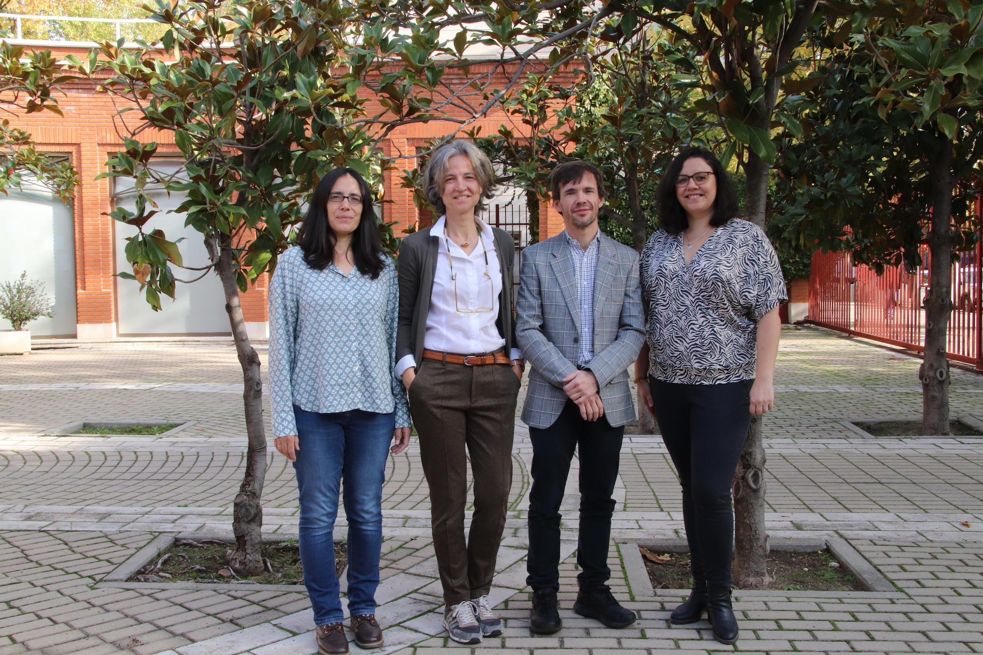
The work has been carried out by the European NanoBright consortium, which includes two Spanish groups: the one led by Manuel Valiente, who heads the CNIO’s Brain Metastasis Group, and the CSIC’s Neuronal Circuits Laboratory of the Cajal Institute, led by Liset Menéndez de la Prida. Both teams have been responsible for the biomedical research at NanoBright, while groups from Italian and French institutions have developed the instrumentation.

Paradigm Shift in Brain Study Techniques
Using light to activate or record brain function is a remarkable achievement, but it is not a new technique. For example, so-called optogenetic techniques make it possible to control the activity of individual neurons with light. However, these methods require the introduction of a gene into the neurons to make them light-sensitive. With the new technology introduced by NanoBright, the brain can be studied without prior alteration, representing a paradigm shift in biomedical research.
The technical name of the method on which the new molecular flashlight is based is vibrational spectroscopy. It works by exploiting a property of light known as the Raman effect: when light interacts with molecules, it scatters differently depending on their chemical composition and structure. This allows for the detection of a unique signal, or spectrum, for each molecule. The spectrum then acts as a molecular signature, providing information about the composition of the illuminated tissue.
“We Can See Any Molecular Change in the Brain Caused by a Pathology or Injury”
“This technology,” explains Manuel Valiente, “allows us to study the brain in its natural state without the need for prior alteration. Moreover, it enables us to analyze any type of brain structure, not just those that have been genetically marked or altered, as was necessary with previous technologies. With vibrational spectroscopy we can see any molecular change in the brain when a pathology is present.”
Raman spectroscopy is already used in neurosurgery, but in a more invasive and less precise manner. “Studies have been conducted on its use during brain tumor surgery in patients,” Valiente notes. “In the operating theatre, once the bulk of the tumor has been removed surgically, a Raman spectroscopy probe can be inserted to assess whether any cancer cells remain in the area. However, this is only done when the brain is already open and the cavity is large enough. These relatively large “molecular flashlights” are incompatible with minimally invasive use in live animal models.”
Minimally Invasive Technique for Analyzing Metastases
The probe developed by the NanoBright consortium is so thin that any damage it may cause when introduced into brain tissue is considered negligible, earning it the designation of “minimally invasive.”
The authors suggest specific applications in Nature Methods. Valiente’s group at CNI, has used the molecular flashlight in experimental models of brain metastases: “As happens with patients, we have observed tumor fronts releasing cells that would escape surgery,” says Valiente. “The difference with existing technology is that we can now perform this analysis in a minimally invasive way, regardless of whether the tumor is superficial or deep.”
For the CNIO team, one current goal is to determine whether the information provided by the probe can “differentiate various oncological entities, such as types of metastases, based on their mutational profiles, by their primary origin or from different types of brain tumors.”
Artificial Intelligence for Identifying Diagnostic Markers
For its part, the Cajal Institute team has used the technique to study the epileptogenic areas around traumatic brain injuries. “We were able to identify different vibrational profiles in the same brain regions prone to epileptic seizures, depending on whether they were associated with a tumor or a trauma. This suggests that the molecular signatures of these areas are affected differently and could be used to distinguish between different pathological entities using automatic classification algorithms, including artificial intelligence,” explains Liset Menéndez de la Prida.
“The integration of vibrational spectroscopy with other modalities for recording brain activity and advanced computational analysis using artificial intelligence will allow us to identify new high-precision diagnostic markers,” concludes the CSIC researcher. “This will facilitate the development of advanced neurotechnology for new biomedical applications.”
Reference: “Vibrational fiber photometry: label-free and reporter-free minimally invasive Raman spectroscopy deep in the mouse brain” by Filippo Pisano, Mariam Masmudi-Martín, Maria Samuela Andriani, Elena Cid, Mohammadrahim Kazemzadeh, Marco Pisanello, Antonio Balena, Liam Collard, Teresa Jurado Parras, Marco Bianco, Patricia Baena, Francesco Tantussi, Marco Grande, Leonardo Sileo, Francesco Gentile, Francesco De Angelis, Massimo De Vittorio, Liset Menendez de la Prida, Manuel Valiente and Ferruccio Pisanello, 31 December 2024, Nature Methods.
DOI: 10.1038/s41592-024-02557-3
Never miss a breakthrough: Join the SciTechDaily newsletter.
Follow us on Google and Google News.