“Dormant” cone photoreceptors continue to drive retinal activity for vision.
New University of California, Los Angeles (UCLA) research in mice suggests that “dormant” cone photoreceptors in the degenerating retina are not dormant at all, but continue to function, producing responses to light and driving retinal activity for vision.
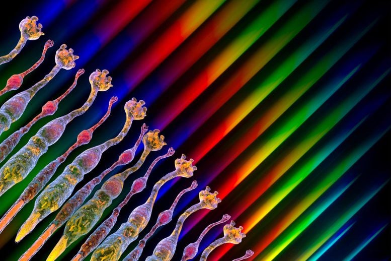
The cells in the retina that produce the visual experience are rods and cones. Rods are active in dim light and cones in daylight. Mutations in rods that cause them to die trigger most inherited retinal degeneration. Cones can remain alive after nearly all the rods die, but they retract key parts of the cells and appear “dormant.”
But while past literature suggested that dormant cells were not functional, and earlier attempts to record from them revealed no light-driven activity, the new study indicates for the first time that the cells are still viable. Furthermore, downstream signals recorded from the retina show that visual processing is not as compromised as may be expected. The authors say their findings demonstrate that therapeutic interventions to protect these cells, or enhance their sensitivity, have the capability to preserve nearly normal daytime vision.
Inner Retina Adaptation
“While the sensitivity of the cones was about 100-1000 fold less than normal, we were surprised to find that that the drop-off in sensitivity for the ganglion cells that project to the brain was much less,” said senior author Alapakkam Sampath, the Grace and Walter Lantz Endowed Chair in Ophthalmology at the UCLA Jules Stein Eye Institute and professor at the David Geffen School of Medicine at UCLA. “It seems that adaptational mechanisms in the inner retina might be trying to minimize the sensitivity difference to preserve robust signaling in the ganglion cells — this is consistent with what we know about the brain. Homeostatic mechanisms that respond to injury and disease typically cover up the deficiency. That is why it is hard to detect neurological problems until the deficiency becomes very severe.”
The study was published recently in the peer-reviewed journal, Current Biology.
The investigators examined membrane properties of cones in mice following the degeneration of rods. The patch clamp recording method is a laboratory technique for studying currents in living cells while controlling the cell’s membrane potential, or membrane voltage. These single cell recordings can establish key features of the cell’s activity, including the presence of specific membrane currents, whether the cell has light responses, and whether they might connect to downstream neurons in the retina. In addition, the investigators used multi-electrode array recordings that establish the activity of all retinal ganglion cells, and that can show the ganglion cell’s ability to respond to visual stimuli that vary in spatial location over time.
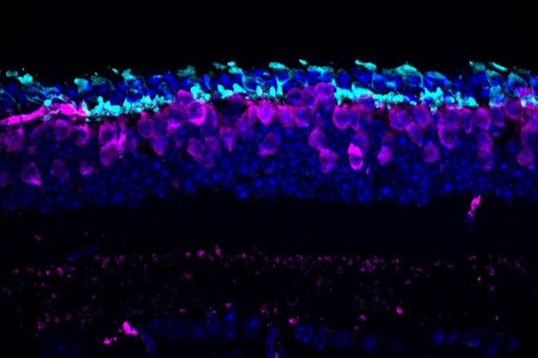
These recordings revealed that the remaining cones in a retina where the rods have mostly degenerated were still functional. Although the anatomic specializations that are responsible for generating the light response — or phototransduction — and the synaptic connection to downstream cells were missing, these functions remained with less sensitivity than normal. These cells still display many of the features of normal cones, including a similar resting membrane potential, a normal synaptic Ca2+ current, and light responses even though they no longer have the part of the cell that was traditionally thought needed for the light response. Furthermore, the ganglion cells retain their ability to respond to visual stimuli with similar spatial and temporal sensitivity.
Implications for Restoring Vision
“These important results may suggest a future path forward for patients with conditions thought to be causing irreversible retinal blindness, as photoreceptor or cone viability in tissue was previously thought to be irreparably damaged,” said Dr. Steven Schwartz, Ahmanson chair in ophthalmology at the David Geffen School of Medicine at UCLA, and professor and Retina Division chief at the UCLA Jules Stein Eye Institute.
The next step for researchers is to establish the extent to which the neuroprotection or enhancement of the dormant cones will allow the rescue of vision in various forms of blindness.
Reference: “Cones and cone pathways remain functional in advanced retinal degeneration” by Erika M. Ellis, Antonio E. Paniagua, Miranda L. Scalabrino, Mishek Thapa, Jay Rathinavelu, Yuekan Jiao, David S. Williams, Greg D. Field, Gordon L. Fain and Alapakkam P. Sampath, 27 March 2023, Current Biology.
DOI: 10.1016/j.cub.2023.03.007
The researchers were supported by grants from the National Eye Institute (R01EY033035, R01EY027442, R01EY27193, R01EY001844, R01EY27193 and EY29817), a fellowship of the UCLA EyeSTAR program of the UCLA Department of Ophthalmology, a BrightFocus Foundation Postdoctoral Fellowship, an unrestricted grant from Research to Prevent Blindness USA to the UCLA Department of Ophthalmology and National Eye Institute Core Grant (P30) EY00311 to the Jules Stein Eye Institute.
The study’s other authors are Dr. Erika Ellis, Antonio Paniagua, Yuekan Jiao, David Williams, Gordon Fain, all of UCLA; and Miranda Scalabrino, Mishek Thapa, Jay Rathinavelu and Greg Field, all of Duke University. The Field laboratory has recently moved to the UCLA Jules Stein Eye Institute. The authors declare no competing interests.
Never miss a breakthrough: Join the SciTechDaily newsletter.
Follow us on Google and Google News.