Researchers have just made the unexpected discovery of a novel organization of muscle fibers in Parophidion vassali, a fish that lives in the Mediterranean Sea and, like many fish, uses specialized muscles to produce sounds. This is an important discovery that could well change our understanding of muscle contraction.
Scientists at the University of Liège, Eric Parmentier and Marc Thiry, have discovered a unique arrangement of muscle fibers in the Mediterranean fish, Parophidion vassali, potentially revolutionizing our understanding of muscle contraction. This unique network-like configuration of myofibrils within the muscle fiber could allow for rapid contractions while retaining strength. Further research is needed to fully understand this novel muscle fiber structure and its functional implications.
The history of the description of skeletal muscles has its origins in the observations made by the Dutch biologist Antoni van Leeuwenhoek, a precursor of cell biology and microbiology who, in an article published in 1712 in the Philosophical Transactions of the Royal Society, reported, thanks to the use of a portable single-lens microscope, the first description of muscle fibers from the whale. In 1840, the anatomist William Bowman provided a more precise description of the muscle, noting: “the existence and arrangement of alternately light and dark lines […] which are of exquisite delicacy and finish.” (See About Muscle Fibers at the bottom of this article.)
Subsequent studies have led to increasingly clear descriptions, including the identification of the different molecules that make up the muscle and an explanation of how it works, in particular the model of muscle contraction proposed by the biophysicist Andrew Huxley in 1957. Over the last 300 years, numerous studies have extended the representation of muscle fiber organization to different taxa. This has shown that the general organization of striated muscle fibers has remained perfectly conserved in all groups of vertebrate animals studied to date.
Variations in Muscle Fiber Composition and Function
However, the proportion of each of these cellular components can vary from one fiber to another, giving these fibers particular contraction properties. For example, a fiber rich in myofibrils with a poorly developed sarcoplasmic reticulum is found in muscles that develop force during contraction. Conversely, fibers low in myofibrils with an abundance of sarcoplasmic reticulum and numerous mitochondria are present in muscles that develop an increased speed of contraction. The fastest muscles are found in the sonic muscles of fish, where certain species produce sounds using muscles that contract at a frequency of between 100 and 300 Hz, i.e. 100 to 300 contraction/relaxation cycles per second.
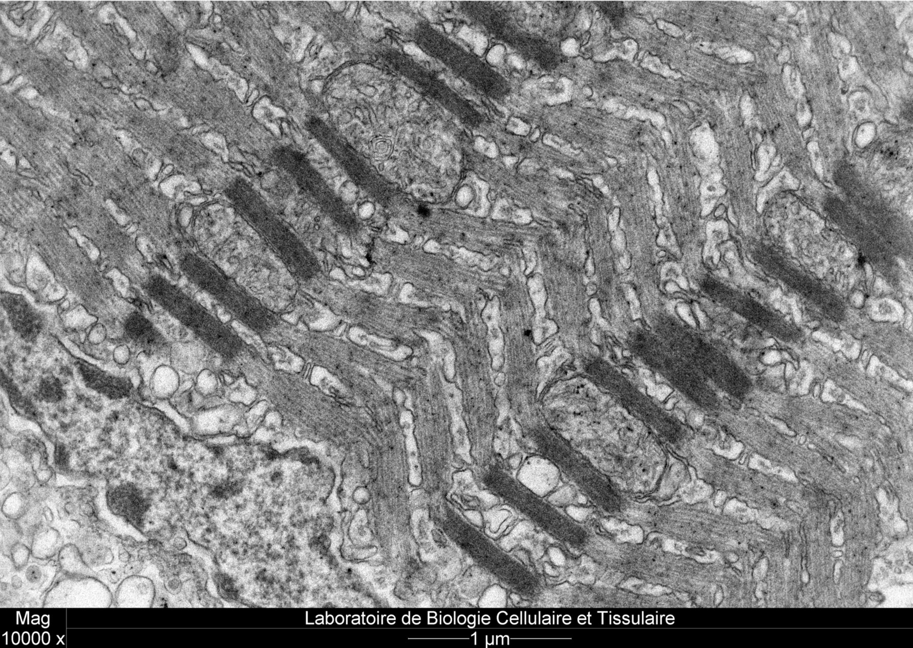
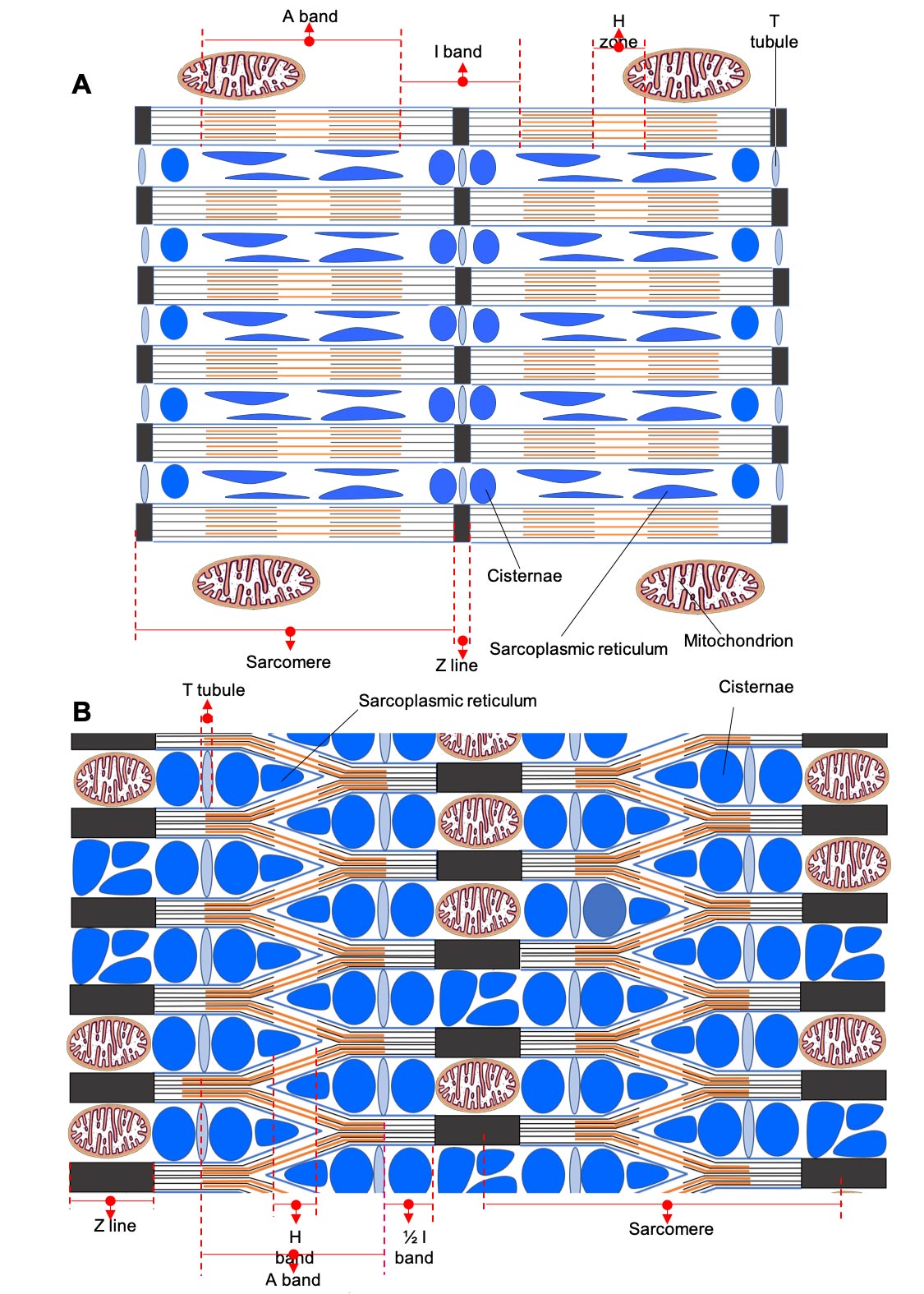
A recent study carried out in collaboration between the Functional and Evolutionary Morphology Laboratory, and the Cell and Tissue Biology Laboratory has revealed a new arrangement of myofibrils within the fibers of a sonic muscle in the fish Parophidion vassali. Rather than being arranged in parallel, the myofibrils form an enormous network within the muscle fiber, explains Prof Eric Parmentier, director of the Functional and Evolutionary Morphology Laboratory at the University of Liège.
Each myofibril subdivides into two branches at each sarcomere, one connecting to the myofibril above and the other connecting to the myofibril below (Figure 1B). This new muscle fiber design could result in a muscle that contracts rapidly while retaining strength. The paucity of myofibrils and the high volume occupied by the sarcoplasmic reticulum are in favor of fibers that contract rapidly.
Myofibril Network Structure
“The network structure of the myofibrils would allow more myosin heads to form cross-bridges with the actin myofilaments, which would increase strength in this fast muscle,” explains Prof Marc Thiry, Director of the Cell and Tissue Biology Laboratory. “In addition, numerous mitochondria unusually arranged within the Z striae (very long in these fibers: 700 nm compared with 70 to 150 nm in a conventional muscle) appear to provide the energy needed to produce long-lasting sounds.”
This new type of organization of striated muscle fibers, never before described in the scientific literature, and which would therefore make it possible to combine muscular strength and speed, requires further studies to understand how it works and to determine whether there are adaptations at the level of the different molecules involved in these muscles.
About Muscle Fibers
Striated skeletal muscle fibers or cells represent the elementary units of voluntary muscles (muscles that enable movements such as locomotion or posture maintenance) in animals. “Each fiber is characterized by numerous contractile elements, actin, and myosin myofilaments, organized in bundles parallel to the muscle fiber’s long axis, called myofibrils.
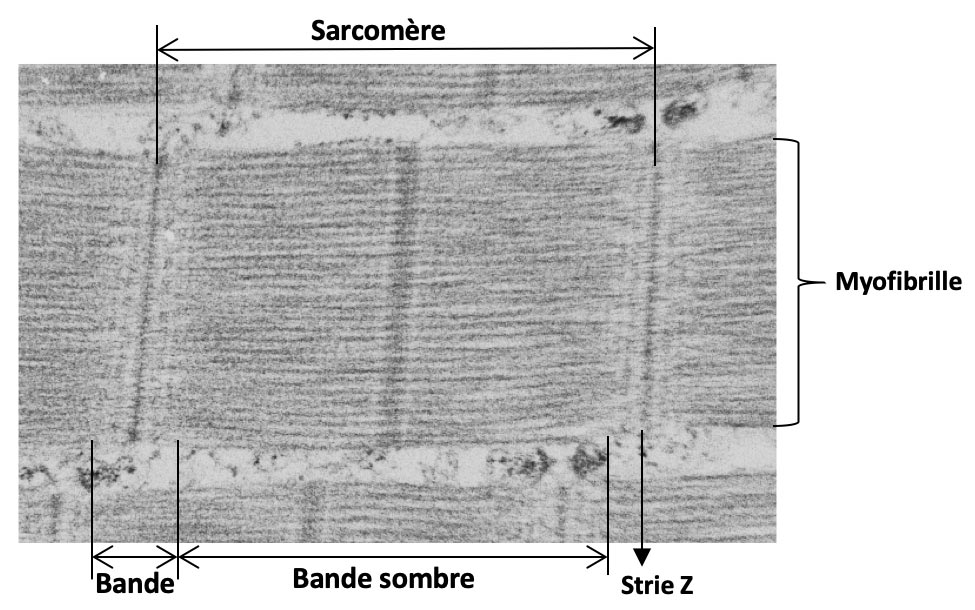
In the longitudinal section under the optical microscope, these fibers appear as a succession of light and dark bands located at the same level for each myofibril, giving the appearance of transverse striation to the muscle fiber. A darker line divides the light band in the middle, known as the Z striation. The portion of the myofibril between two Z striae is called a sarcomere and represents the contractile unit of the myofibril. Each myofibril is therefore made up of many sarcomeres placed end to end.
The myofibrils occupy a large cell volume and are surrounded by cisternae of the smooth endoplasmic reticulum (or sarcoplasmic reticulum), which stores the calcium essential for muscle contraction. In addition, mitochondria are located close to the myofibrils; they are the main source of ATP providing energy for muscle contraction.
Never miss a breakthrough: Join the SciTechDaily newsletter.
Follow us on Google and Google News.