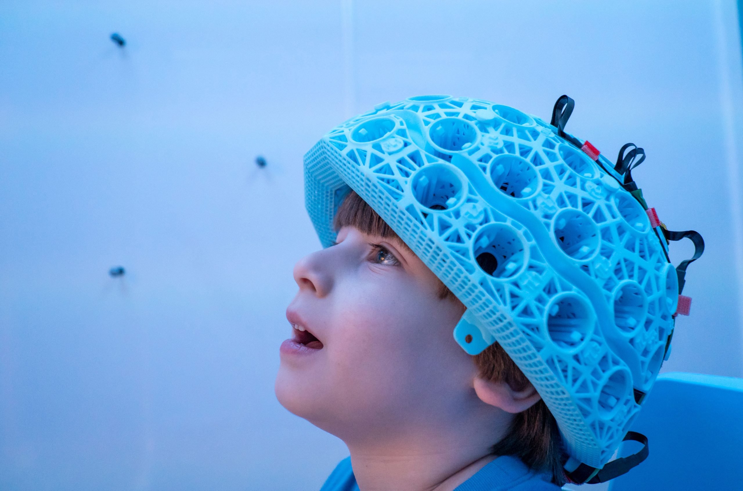
Child wearing one of the MEG-OPM helmet style brain scanners. Credit: University of Nottingham
Recent research using wearable brain scanners offers new insights into children’s brain development, tracking how critical milestones are linked to brain activity and exploring the foundations of neurodevelopmental conditions like autism.
New research has provided the most detailed view yet of the developing brains of young children by using a wearable brain scanner to chart electrical activity. This study paves the way for new methods to monitor how essential developmental milestones, such as walking and talking, are supported by evolving brain functions, and to explore the origins of neurodevelopmental conditions like autism.
The research team used a novel design of magnetoencephalography (MEG) scanner to measure brain electrophysiology in children as young as two. Scientists from the University of Nottingham’s School of Physics and Astronomy led the study, and the findings were published in the journal eLife.
Brain cells operate and communicate by producing electrical currents. These currents generate tiny magnetic fields that can be detected outside the head. Researchers used their novel system to measure these fields, and mathematical modeling to turn those fields into high-fidelity images showing, millisecond-by-millisecond, which parts of the brain are engaged when we undertake tasks.
Innovations in Wearable Brain Scanners
The wearable brain scanner is based on quantum technology, and uses LEGO-brick-sized sensors – called optically pumped magnetometers (OPMs) – which are incorporated into a lightweight helmet to measure the fields generated by brain activity. The unique design means the system can be adapted to fit any age group, from toddlers to adults. Sensors can be placed much closer to the head, enhancing data quality. The system also allows people to move whilst wearing it, making it ideal for scanning children who find it hard to keep still in conventional scanners.
27 children (aged 2-13 years) and 26 adults (aged 21-34 years) took part in the study, which examined a fundamental component of brain function called ‘neural oscillations’ (or brain waves). Different areas of the brain are responsible for different aspects of behavior and neural oscillations promote communication between these regions. The research team measured how this connectivity changes as we grow up, and how our brains use short, punctate bursts of electrophysiological activity to inhibit networks of brain regions, and consequently to control how we attend to incoming sensory stimuli.
Comments from the Research Leaders
The work was jointly led by Dr. Lukas Rier, and Dr. Natalie Rhodes from the University of Nottingham’s School of Physics and Astronomy. Dr. Rier said: “The wearable system has opened up new opportunities to study and understand children’s brains at much younger ages than was previously possible with MEG. There are important reasons for moving to younger participants: from a neuroscientific viewpoint, many critical milestones in development occur in the first few years (even months) of life. If we can use our technology to measure the brain activities that underpin these developmental milestones, this would offer a new understanding of brain function.”
The research, which was funded by the Engineering and Physics Research Council (EPSRC), included academic collaborators from SickKids Hospital in Toronto, Canada, and industry partners from US-based atomic device company QuSpin and Nottingham-based company Cerca Magnetics Limited.
Dr. Rhodes was a University of Nottingham undergraduate student in Physics, and a postgraduate student when the work was carried out. She has now moved to a postdoctoral position in Toronto, and explains: “This study is the first of its kind using wearable MEG technology and provides a platform to launch new clinical research in childhood disorders. This means that we can begin to explore not only healthy brain development, but also the neural substrates that underlie atypical development in children.”
World-renowned neuroscientist Dr. Margot Taylor – also an author on the paper – is leading research into autism in Toronto. She said: “Our work is dedicated to studying brain function in young children with and without autism. This study is the first to demonstrate that we can track brain development from a very young age. This is hugely exciting for possible translation to clinical research and work such as this helps us understand how autism develops.”
Reference: “Tracking the neurodevelopmental trajectory of beta band oscillations with optically pumped magnetometer-based magnetoencephalography” by Lukas Rier, Natalie Rhodes, Daisie O Pakenham, Elena Boto, Niall Holmes, Ryan M Hill, Gonzalo Reina Rivero, Vishal Shah, Cody Doyle, James Osborne, Richard W Bowtell, Margot Taylor and Matthew J Brookes, 4 June 2024, eLife.
DOI: doi:10.7554/eLife.94561
The University launched a spin-out company Cerca Magnetics in 2020 to commercialise OPM-MEG scanners and related technologies. The wearable system has been installed in a number of high-profile research institutions across the globe, including SickKids hospital in Toronto. The research teams in both institutions are now working together to expand the amount of neurodevelopmental data, on both healthy and atypical brain function


Isn’t that, like, child abuse?