
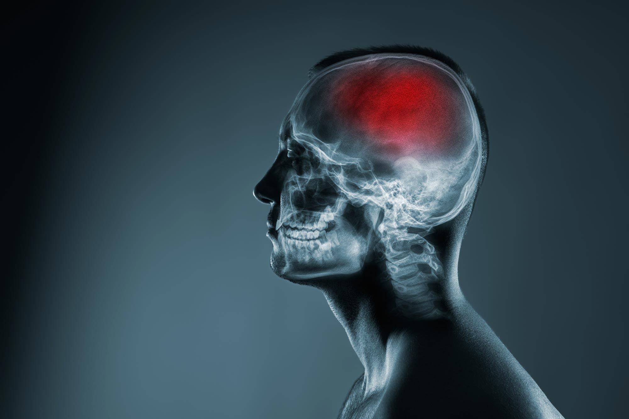
An injectable biomaterial turned stroke-damaged areas into hubs of repair, helping mouse brains grow new blood vessels and nerve fibers while restoring near-normal movement.
A stroke can leave behind more than damaged brain cells. In severe cases, it creates an empty cavity where living tissue once carried signals, supplied blood, and controlled movement. Duke University researchers are now testing an injectable material designed to turn that biological void into a place where repair can begin.
In mice, the treatment drew immune cells into the stroke cavity and helped organize them into a coordinated healing response. New blood vessels spread through the injured area, nerve fibers became more abundant, and the animals regained motor abilities that approached those of healthy mice.
The findings were published in Cell Biomaterials. The material was injected directly into the damaged region five days after the stroke, meaning it was tested as a repair strategy rather than an emergency treatment.
Why Stroke Damage Is So Hard to Repair
Most strokes occur when a clot cuts off blood flow to part of the brain. Clot-dissolving drugs and procedures that physically remove the blockage can save threatened tissue when delivered quickly. Once brain cells have died, however, restoring circulation cannot bring them back.
A major ischemic stroke may destroy enough tissue to leave a fluid-filled cavity. Rehabilitation can train surviving brain networks to take on new roles, but medicine currently has no established way to reconstruct the missing region itself.
“Once brain tissue has been lost, restoring blood flow is no longer enough,” said Tatiana Segura, the Robert Plonsey Distinguished Professor of Biomedical Engineering at Duke. “Our goal is to engineer the injured space so that immune, vascular, and neural repair processes can begin to work together.”

An Injectable Scaffold for Brain Repair
Rather than trying to manufacture replacement brain tissue, Segura’s team developed a temporary framework that encourages the body to do more of the rebuilding itself.
The treatment is based on MAPS, or microporous annealed particle scaffolds. These injectable materials are assembled from tiny hydrogel particles that connect after delivery while leaving open spaces between them. Unlike a solid gel, the porous structure gives cells room to enter, move, and form new tissue. Microporous scaffolds can support cellular infiltration and blood vessel growth without waiting for the entire material to break down first.
The Duke team had previously investigated similar materials for stroke repair. In the new work, the researchers added biological instructions intended to shape the immune response inside the scaffold.
Astrocyte Signals Guide the Healing Response
Those instructions came from astrocytes, star-shaped cells that support neurons, help regulate the brain’s environment, and react rapidly to injury. Astrocytes communicate partly by releasing extracellular vesicles, or EVs. These nanoscale packages transport proteins, lipids, and genetic material between cells.
Researchers grew astrocytes in the laboratory and exposed them to different signaling molecules. They then collected the EVs produced under those conditions and tested whether the packages could attract immune cells and encourage tissue repair.
Simply releasing EVs into the damaged brain would allow many of them to disperse. To keep the signals where they were needed, the researchers chemically attached the vesicles to the hydrogel particles.
Turning the Scaffold Into a Signaling Hub
This design transformed the scaffold into more than a physical support. It became a localized signaling hub where incoming cells could repeatedly encounter molecular instructions.
“We are not simply placing a material into the brain,” Segura said. “We are engineering a local environment that can coordinate several parts of the repair response.”
EVs produced after astrocytes were exposed to IL-4 and C1q generated the strongest results. The combination attracted macrophages and a surprisingly persistent population of neutrophils into the stroke cavity.
Immune Cells Take on a Surprising Role
Neutrophils are among the immune system’s fastest responders. After a stroke, they are often associated with inflammation and additional tissue damage, especially during the early phase of injury. Yet immune cells do not always have a single fixed role. Their behavior can change depending on timing, location, and the molecular signals surrounding them.
Inside the engineered scaffold, neutrophils appeared to become part of the repair process rather than merely contributing to destruction.
The researchers tested that possibility by depleting the immune-cell population rich in neutrophils. Blood vessel formation dropped sharply, and the scaffold underwent far less remodeling. The experiment showed that these cells were not simply present at the injury site. They were helping drive the response.
“This result changes how we think about neutrophils after stroke,” said Shangjing Xin, lead scientist of the study and a postdoctoral fellow in the Segura Laboratory. “Their role appears to depend on when they arrive, where they are located, and the signals they receive from their surroundings. Our study demonstrates a potential engineering strategy to recruit and retain these cells at the right time.”
New Blood Vessels and Nerve Fibers Emerge
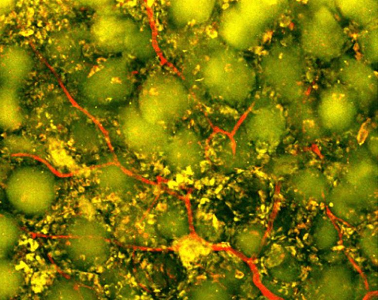
The treatment produced visible changes throughout the damaged region. Blood vessels grew across the cavity, potentially creating the circulation needed to support living tissue. Researchers also detected more axonal fibers within and around the injury. Axons are the long projections neurons use to carry electrical signals to other cells.
Those biological changes were accompanied by improved movement.
During a grid-walking test, scientists measured how often the mice misplaced a front paw while crossing an uneven surface. Animals treated with the optimized scaffold made fewer errors over time. By eight weeks, their performance could not be statistically distinguished from that of healthy control mice, and the improvement continued through the end of the study.
The scaffold itself proved essential. When researchers delivered the EVs without MAPS, they did not observe comparable blood vessel growth. The result suggests that the treatment depended on both components: the biological messages carried by the vesicles and the porous structure that concentrated those messages while giving cells space to organize.
Toward a Scalable Human Stroke Therapy
The study relied on EVs collected from primary rat astrocytes, which would not be a practical source for a widely available human therapy.
Segura’s laboratory is now exploring astrocytes made from human-induced pluripotent stem cells. These cells can be produced from reprogrammed adult cells and expanded in the laboratory, potentially offering a more scalable and clinically relevant source of EVs. Researchers may also be able to adjust the conditions under which the astrocytes grow to better control the messages their vesicles carry.
“You do not restore an ecosystem simply by containing the initial damage,” Segura said. “You have to create the conditions that allow life to return. That is how we think about the stroke cavity. The material is not intended to reproduce the brain itself but to create an environment where the body’s own cells can enter, communicate, and participate in rebuilding vascularized tissue.”
Reference: “IL-4/C1q activated astrocyte-derived extracellular vesicles promote stroke infarct recovery by recruiting peripheral leukocytes” by Shangjing Xin, Lucy Zhang, Nhi V. Phan, Mengying An, Ligen Shi, S. Thomas Carmichael and Tatiana Segura, 21 July 2026, Cell Biomaterials.
DOI: 10.1016/j.celbio.2026.100543
Never miss a breakthrough: Join the SciTechDaily newsletter.
Follow us on Google and Google News.
3 Comments
I am very happy to read all the brain diseases and plus insomnia. These are very knowledgeable and I love to get new treatments
My husband would be a candidate for this research – please let us know if there is a clinical trial in the works
Mora Therapy is also known to promote the healing process. Check out the NIH National Library of Medicine research articles on how they treated some of the most chronic debilitating pathologies globally. Pmc article numbers are: ncbi.nlm.nih.gov/pmc/articles/PMC3845545/
then PMC9595267
then PMC8892610
For more info you can request it at Bioresonance.Biofeedback at yahoo.
They’ll send other information you can look up and read about like FSM (Frequency Specific Microcurrent) used in the NFL and one hospital in the usa.