Researchers have developed a method using viruses to track neuronal development in frogs, shedding light on the evolution of vertebrate nervous systems and offering comparative insights with mammals.
Although viruses are typically associated with illnesses, not all viruses are harmful or cause disease. Some are instrumental in therapeutic treatments and vaccinations. In scientific research, viruses are often used to infect certain cells, genetically modify them, or visualize neurons in the organism’s central nervous system (CNS)—the command center made up of the brain, spinal cord, and nerves.
The highlighting process has now been successfully applied to amphibians, which are crucial for understanding the brain and spinal cord of tetrapods—four-limbed animals, including humans. This has been shown in a new study by an international EDGE consortium jointly led by the Sweeney Lab at the Institute of Science and Technology Austria (ISTA) and the Tosches Lab at Columbia University.
The researchers developed a new method that uses adeno-associated viruses (AAVs) to track a frog’s nervous system throughout its metamorphosis—a developmental transition from the early tadpole stages to its adult form. This groundbreaking research, recently published in Developmental Cell, can help usher amphibian neurobiology into a new era.

From Swimming to Walking: Studying Metamorphosis
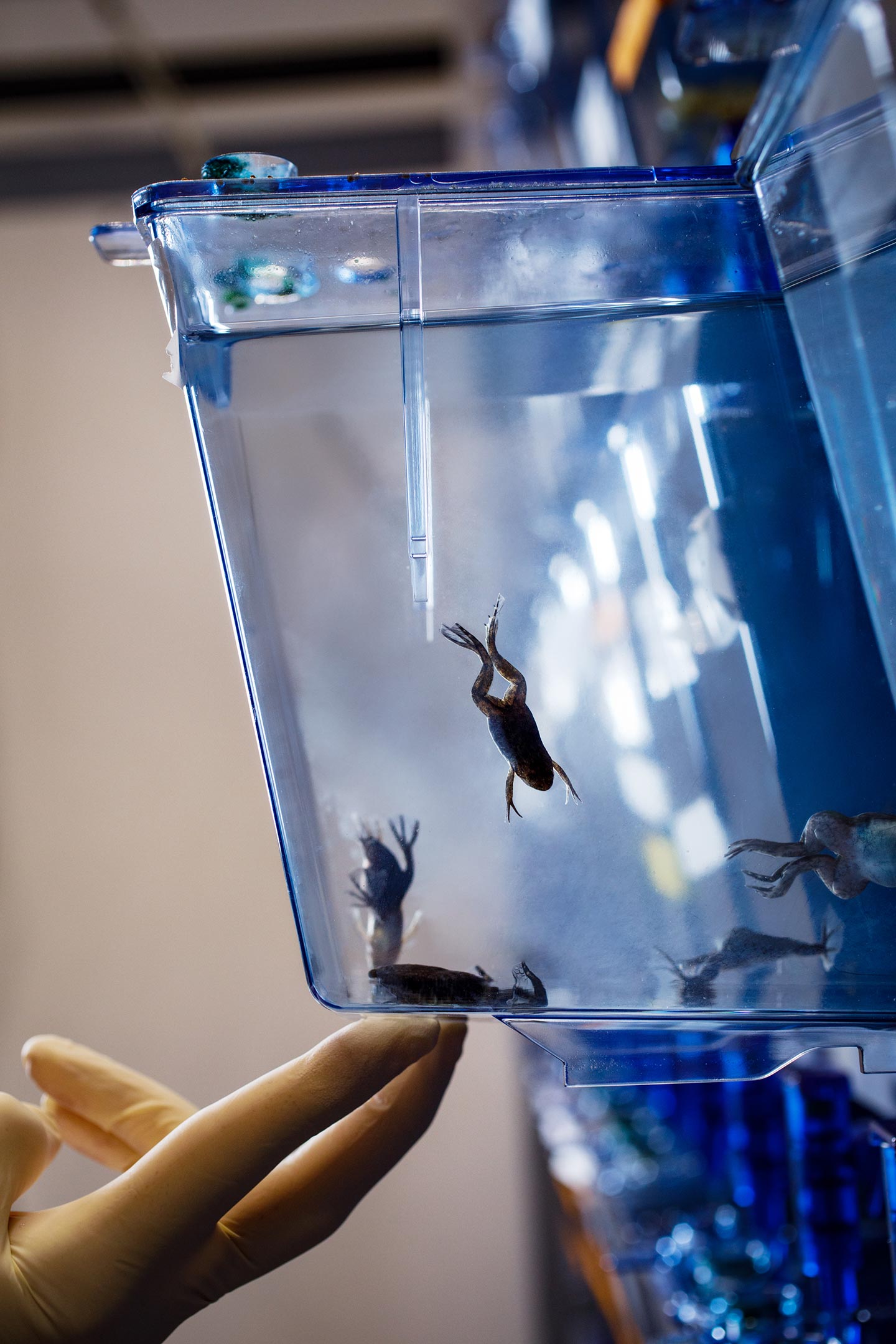
David Vijatovic and Lora Sweeney enter a laboratory full of water tanks. Vijatovic taps on one of them. Inside, a small mottled greenish-brown African clawed frog (Xenopus laevis) appears. Its limbs are prominent, gracefully maneuvering and gripping its surroundings. In another tank, tadpoles are swirling around using simple swimming motions. It is remarkable to think that one transforms into the other.
“Frogs undergo metamorphosis,” Sweeney says, “making them a great model organism for studying the transition between two movement modes—swimming and walking.” A frog’s development spans over 12 to 16 weeks, giving scientists time to study each stage. During these weeks, a frog embryo develops into a young tadpole, a tadpole with two legs, and a young froglet with four legs before reaching the adult stage. “By looking at the several stages of development, we can investigate these locomotive behaviors and the underlying changes in the nervous system,” Vijatovic adds.
Decoding Frog Neural Circuits
An organism’s nervous system is called the neural circuit because it resembles an electrical circuit. “Nerve cells (neurons) are connected to other neurons, transmitting electrical information. How we behave, what we sense, and how we interact with the world are the product of the way our neurons communicate with each other within these circuits,” explains Sweeney. The critical piece is how the circuit is wired. We know that neurons are connected, but which neuron connects to which? Which other cells does a single cell talk to, and what messages does it convey?

To know more about this wiring, researchers have been using viruses, which have proven to be a powerful tool. Adeno-associated viruses (AAVs) are ideal in that regard. They are non-pathogenic and can infect a wide range of cell types, including neurons. AAVs can be modified to glow in bright green fluorescent colors under the microscope as they travel along neurons, whether in retrograde (backward, from the synapse toward the cell body) or anterograde (forward, from the cell body toward the synapse). In other words, AAVs can be used to illuminate the neural circuit from the broadcasting end to the receiving end or vice-versa.
“This is a common technique used in neuroscience, especially in well-studied organisms like mice. For amphibians, it was thought that it could not be done,” says Vijatovic. That was the general belief until now.
Overcoming Barriers With Collaboration
To make AAV labeling work in amphibians, Sweeney and Vijatovic joined forces with an international team of scientists from Maria Tosches’ group at Columbia University, where the study’s other two co-first authors, Eliza Jaeger and Astrid Deryckere, are based. The consortium also included researchers from Tel Aviv University, the University of Utah, the Scripps Research Institute, and the California Institute of Technology. The researchers put their heads together, drew expertise from each other, visited conferences, had countless Zoom calls, and came up with different perspectives and ideas. “When you start researching an organism that is not yet well understood, it is great to have a community where you can share information,” says Sweeney.

They screened existing AAVs to find what was suitable for amphibians and optimized the infecting strategy, eventually developing a “how-to guide” for frogs and newts. Vijatovic summarizes his PhD journey, “We started with young tadpoles, made our way to older tadpoles, and finally moved to juvenile and then adult frogs as well as adult newts. We tailored the tool to each life stage.”
Insights into Human Neuroscience From Amphibians
With this new technique, the scientists managed to apply AAVs to trace neuron connections in amphibians. This will help them find out more about how the amphibian brain compares to that of mammals. Besides that, the new approach also opens doors to further analyzing neuronal development. With some of the screened AAV variants, the researchers can label progenitor cells at a specific point in time during the circuit’s development and follow them to see what neurons they become. “This way, we can resolve the whole circuit by its development, see how it changes over time, and how the whole nervous system is built,” Sweeney says.
Although amphibians and mammals last shared a common ancestor about 360 million years ago, they share common traits. “By comparing the details of a frog’s nervous system to a human’s, we can see what we don’t have and what we have,” Sweeney continues. This knowledge can help us understand how the human nervous system became specialized over time. “The better we understand the basic building blocks of the nervous system, the more we understand how we can replace them during disease and injury.”
Reference: “Adeno-associated viral tools to trace neural development and connectivity across amphibians” by Eliza C.B. Jaeger, David Vijatovic, Astrid Deryckere, Nikol Zorin, Akemi L. Nguyen, Georgiy Ivanian, Jamie Woych, Rebecca C. Arnold, Alonso Ortega Gurrola, Arik Shvartsman, Francesca Barbieri, Florina A. Toma, Hollis T. Cline, Timothy F. Shay, Darcy B. Kelley, Ayako Yamaguchi, Mark Shein-Idelson, Maria Antonietta Tosches and Lora B. Sweeney, 26 November 2024, Developmental Cell.
DOI: 10.1016/j.devcel.2024.10.025
Never miss a breakthrough: Join the SciTechDaily newsletter.
Follow us on Google and Google News.