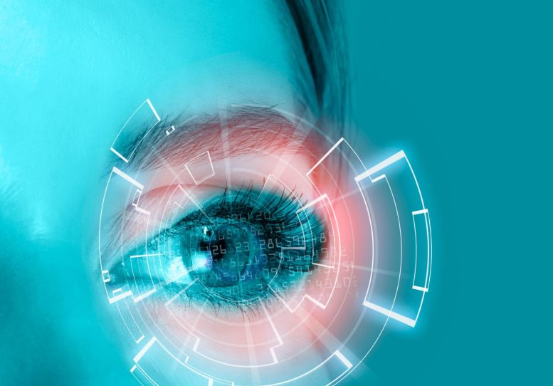
An AI-powered proteomic clock from eye fluid reveals disease-driven aging and may enable early Parkinson’s detection.
A team of researchers has mapped almost 6,000 proteins from different cell types within the eye by analyzing tiny drops of eye fluid that are routinely removed during surgery. In a study recently published in the journal Cell, the researchers used an AI model to create a “proteomic clock” from this data that can predict a healthy person’s age based on their protein profile.
The clock revealed that diseases such as diabetic retinopathy and uveitis cause accelerated aging within specific cell types. Surprisingly, the researchers also detected proteins associated with Parkinson’s disease within eye fluid, which they say could offer a pathway to earlier Parkinson’s diagnoses.
Eye as a Window to Diseases
“What’s amazing about the eye is we can look inside and see diseases happening in real-time,” says senior author Vinit Mahajan, a surgeon and professor of ophthalmology at Stanford University. “Our primary focus was to connect those anatomical changes to what’s happening at the molecular level inside the eyes of our patients.”
The eye is a difficult organ to sample in living patients because, like the brain, it is non-regenerative, and taking a tissue biopsy would cause irreparable damage. An alternative method is to use liquid biopsies—samples of fluid taken from near the cells or tissues of interest.
Though liquid biopsies can provide a snapshot of what proteins are present in the region of interest, they have thus far been limited in their ability to measure large numbers of proteins within the small volumes of fluid, and they are also unable to provide information on which cells produced which proteins, which is important for diagnosing and treating diseases.
Advanced Protein Mapping and Findings
To map protein production by different types of cells within the eye, Mahajan’s team used a high-resolution method to characterize proteins in 120 liquid biopsies taken from the aqueous or vitreous humor of patients undergoing eye surgery. Altogether, they identified 5,953 proteins—ten times the number of proteins previously characterized in similar studies. Using a software tool they created called TEMPO, the researchers were able to trace each protein back to specific cell types.
To investigate the relationship between disease and molecular aging, the researchers built an AI machine-learning model that can predict the molecular age of the eye based on a subset of 26 proteins. The model was able to accurately predict the age of healthy eyes but showed that diseases were associated with significant molecular aging. For diabetic retinopathy, the degree of aging increased with disease progression and this aging was accelerated by as much as 30 years for individuals with severe (proliferative) diabetic retinopathy. These signs of aging were sometimes observable before the patient displayed clinical symptoms of the underlying disease and lingered in patients who had been successfully treated.
The researchers also detected several proteins that are associated with Parkinson’s disease. These proteins are usually identified postmortem and current diagnostic methods aren’t capable of testing for them, which is one reason Parkinson’s diagnoses are so difficult. Screening for these markers in eye fluid could enable earlier diagnosis of Parkinson’s disease and later therapeutic monitoring.
Implications and Future Directions
The authors say that these results suggest that aging may be organ- or even cell-specific, which could yield advances in precision medicine and clinical trial design. “These findings demonstrate that our organs are aging at different rates,” says first author and ophthalmologist Julian Wolf of Stanford University. “The use of targeted anti-aging drugs could be the next step in preventative, precision medicine.”
“If we’re going to use molecular therapies, we should be characterizing the molecules in our patients,” says Mahajan. “I think reclassifying patients based on their molecular patterns and which cells are being affected can really improve clinical trials, drug selection, and drug outcomes.”
Next, the researchers plan to characterize samples from a larger number of patients and a broader range of eye diseases. They also say that their method could be used to characterize other difficult-to-sample tissues. For example, liquid biopsies of cerebrospinal fluid could be used to study or diagnose the brain, synovial fluid could be used to study joints, and urine could be used to study the kidneys.
Reference: “Liquid-biopsy proteomics combined with AI identifies cellular drivers of eye aging and disease in vivo” by Julian Wolf, Ditte K. Rasmussen, Young Joo Sun, Jennifer T. Vu, Elena Wang, Camilo Espinosa, Fabio Bigini, Robert T. Chang, Artis A. Montague, Peter H. Tang, Prithvi Mruthyunjaya, Nima Aghaeepour, Antoine Dufour, Alexander G. Bassuk and Vinit B. Mahajan, 19 October 2023, Cell.
DOI: 10.1016/j.cell.2023.09.012
This research was supported by the National Institutes of Health, Stanford University, Research to Prevent Blindness, the VitreoRetinal Surgery Foundation, the Lundbeck Foundation, and the BrightFocus Foundation.
Never miss a breakthrough: Join the SciTechDaily newsletter.
Follow us on Google and Google News.
2 Comments
Very very good research using this creative approach to analyse should be widely used in diagnosing, planing a path of solution for multiple health problems and possibly creating a method of prevention in alife long detection starting a birth, simply remarkable work.
Leave no stone unturned