Close Menu
    Facebook X (Twitter) Instagram
    SciTechDaily
    • Biology
    • Chemistry
    • Earth
    • Health
    • Physics
    • Science
    • Space
    • Technology
    Facebook X (Twitter) Pinterest YouTube RSS
    SciTechDaily
    Home»Health»New Study: Light Therapy Enhances Brain Connectivity After Injury
    Health

    New Study: Light Therapy Enhances Brain Connectivity After Injury

    By Radiological Society of North AmericaJune 4, 20241 Comment5 Mins Read
    Facebook Twitter Pinterest Telegram LinkedIn WhatsApp Email Reddit
    Share
    Facebook Twitter LinkedIn Pinterest Telegram Email Reddit
    Brain Injury Stroke
    Recent research highlights the potential benefits of low-level light therapy in enhancing brain connectivity after moderate traumatic brain injuries, though its long-term effects and clinical implications remain to be fully understood.

    Low-level light therapy may enhance brain connectivity in patients with moderate traumatic brain injuries.

    According to a study published on May 28 in Radiology, a journal of the Radiological Society of North America (RSNA), low-level light therapy seems to influence healing in the brains of individuals who have experienced significant brain injuries.

    Lights of different wavelengths have been studied for years for their wound-healing properties. Researchers at Massachusetts General Hospital (MGH) conducted low-level light therapy on 38 patients who had suffered moderate traumatic brain injury, an injury to the head serious enough to alter cognition and/or be visible on a brain scan. Patients received light therapy within 72 hours of their injuries through a helmet that emits near-infrared light.

    “The skull is quite transparent to near-infrared light,” said study co-lead author Rajiv Gupta, M.D., Ph.D., from the Department of Radiology at MGH. “Once you put the helmet on, your whole brain is bathing in this light.”

    Measuring Brain Connectivity Changes

    The researchers used an imaging technique called functional MRI to gauge the effects of the light therapy. They focused on the brain’s resting-state functional connectivity, the communication between brain regions that occurs when a person is at rest and not engaged in a specific task. The researchers compared MRI results during three recovery phases: the acute phase of within one week after injury, the subacute phase of two to three weeks post-injury, and the late-subacute phase of three months after injury.

    Of the 38 patients in the trial, 21 did not receive light therapy while wearing the helmet. This was done to serve as a control to minimize bias due to patient characteristics and to avoid potential placebo effects.

    Functional MRI Brain Maps of Resting State Functional Connectivity
    Functional MRI brain maps of resting-state functional connectivity in representative age- and sex-matched participants. (A, B) Axial (top) and coronal (bottom) views show whole-brain connectivity, with the seed at the left (L) superior frontal region, in a 36-year-old female participant in the low-level light therapy (LLLT) treatment group (A) and a 38-year-old female participant in the sham treatment group (B) during the acute, subacute, and late-subacute phases (columns, from left to right, in both A and B) of traumatic brain injury recovery. (C) Axial (top left), coronal (bottom left), and sagittal (right) views in a 38-year-old female control participant are shown for comparison; the solid green circle in the sagittal view indicates the location of the left superior frontal seed region. The color bar indicates that brain regions with warm colors (red, orange, yellow) show resting-state fluctuations that have significant positive correlation (r of 0 to 1) with those of the left superior frontal region, and brain regions with cold colors (blue) show resting-state fluctuations that have significant negative correlation (r of −1 to 0) with those of the left superior frontal region. Brain regions that have functional connectivity with the left superior frontal seed in the LLLT-treated participant (arrowheads in A) but not in the sham-treated participant (arrowheads in B) are shown. The arrow in A additionally shows brain regions with positive correlation with the seed in the LLLT-treated participant, but negative correlation with the seed in the sham-treated participant (arrow in B). Credit: Radiological Society of North America (RSNA)

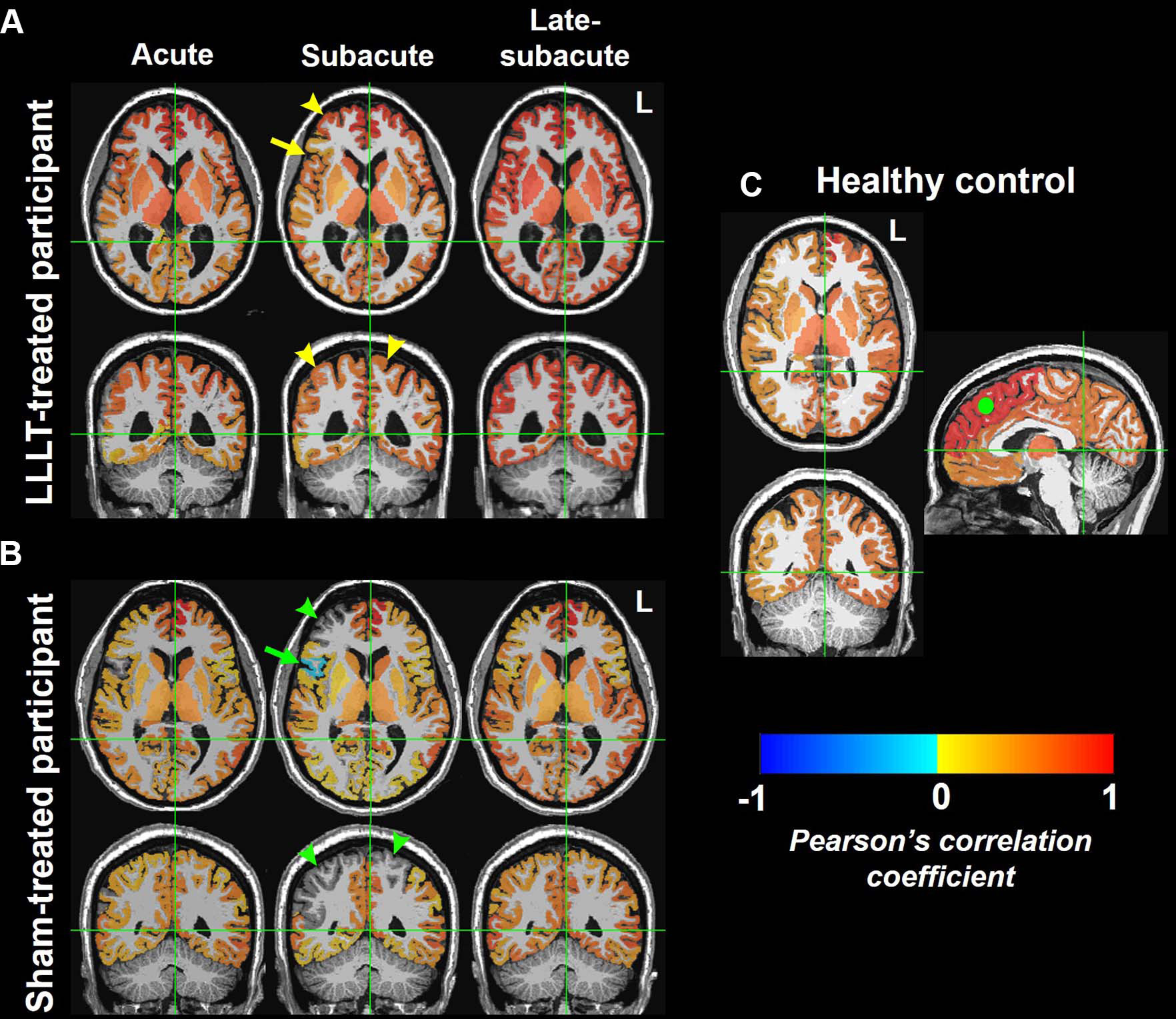
    Patients who received low-level light therapy showed a greater change in resting-state connectivity in seven brain region pairs during the acute-to-subacute recovery phase compared to the control participants.

    “There was increased connectivity in those receiving light treatment, primarily within the first two weeks,” said study coauthor Nathaniel Mercaldo, Ph.D., a statistician with MGH. “We were unable to detect differences in connectivity between the two treatment groups long term, so although the treatment appears to increase the brain connectivity initially, its long-term effects are still to be determined.”

    Mechanisms and Future Research

    The precise mechanism of the light therapy’s effects on the brain is also still to be determined. Previous research points to the alteration of an enzyme in the cell’s mitochondria (often referred to as the “powerhouse” of a cell), Dr. Gupta said. This leads to more production of adenosine triphosphate, a molecule that stores and transfers energy in the cells. Light therapy has also been linked with blood vessel dilation and anti-inflammatory effects.

    “There is still a lot of work to be done to understand the exact physiological mechanism behind these effects,” said study coauthor Suk-tak Chan, Ph.D., a biomedical engineer at MGH.

    While connectivity increased for the light therapy-treated patients during the acute to subacute phases, there was no evidence of a difference in clinical outcomes between the treated and control participants. Additional studies with larger cohorts of patients and correlative imaging beyond three months may help determine the therapeutic role of light in traumatic brain injury.

    The researchers expect the role of light therapy to expand as more study results come in. The 810-nanometer-wavelength light used in the study is already employed in various therapeutic applications. It’s safe, easy to administer, and does not require surgery or medications. The helmet’s portability means it can be delivered in settings outside of the hospital. It may have applications in treating many other neurological conditions, according to Dr. Gupta.

    “There are lots of disorders of connectivity, mostly in psychiatry, where this intervention may have a role,” he said. “PTSD, depression, autism: these are all promising areas for light therapy.”

    Reference: “Effects of Low-Level Light Therapy on Resting-State Connectivity Following Moderate Traumatic Brain Injury: Secondary Analyses of a Double-blinded Placebo-controlled Study” by Suk-tak Chan, Nathaniel Mercaldo, Maria G. Figueiro Longo, Jonathan Welt, Arman Avesta, Jarone Lee, Michael H. Lev, Eva-Maria Ratai, Michael R. Wenke, Blair A. Parry, Lynn Drake, Richard R. Anderson, Terry Rauch, Ramon Diaz-Arrastia, Kenneth K. Kwong, Michael Hamblin, Benjamin J. Vakoc and Rajiv Gupta, 28 May 2024, Radiology.
    DOI: 10.1148/radiol.230999

    Never miss a breakthrough: Join the SciTechDaily newsletter.
    Follow us on Google and Google News.

    Brain MRI Neurology Radiological Society of North America
    Share. Facebook Twitter Pinterest LinkedIn Email Reddit

    Related Articles

    Scientists Discover Early Alzheimer’s Warning Sign Hiding in Routine Brain Scans

    Fighters’ Brains Show a Surprising Cleanup Surge Before Dangerous Collapse

    Soccer Players Beware – New Research Links Heading to Significant Decline in Brain Function

    Can This Medication Reverse Multiple Sclerosis? Brain Biomarker Shows It Can

    Artificial Intelligence Classifies Brain Tumors With Single MRI Scan

    Noninvasive Arterial Spin Labeling MRI Detects Evidence of Cognitive Decline Before Symptoms Appear

    Disrupted Sleep Patterns Linked to Alzheimer’s

    Custom Tailored Brain Cancer Vaccine Proves Effective

    Size Does Matter When Determining Alzheimer’s Risk

    1 Comment

    1. kamir bouchareb st on June 5, 2024 3:05 am

      goooooood

      Reply
    Leave A Reply Cancel Reply

    • Facebook
    • Twitter
    • Pinterest
    • YouTube

    Don't Miss a Discovery

    Subscribe for the Latest in Science & Tech!

    Trending News

    Your Blood Pressure Reading Could Be Wrong Because of One Simple Mistake

    Astronomers Stunned by Ancient Galaxy With No Spin

    Physicists May Be on the Verge of Discovering “New Physics” at CERN

    Scientists Solve 320-Million-Year Mystery of Reptile Skin Armor

    Scientists Say This Daily Walking Habit May Be the Secret to Keeping Weight Off After Dieting

    New Therapy Rewires the Brain To Restore Joy in Depression Patients

    Giant Squid Detected off Western Australia in Stunning Deep-Sea Discovery

    Popular Sugar-Free Sweetener Linked to Liver Disease, Study Warns

    Follow SciTechDaily
    • Facebook
    • Twitter
    • YouTube
    • Pinterest
    • Newsletter
    • RSS
    SciTech News
    • Biology News
    • Chemistry News
    • Earth News
    • Health News
    • Physics News
    • Science News
    • Space News
    • Technology News
    Recent Posts
    • New Stroke Study Challenges Decades-Old Medical Beliefs
    • These Simple Plant Foods Are Linked to Lower Blood Pressure
    • Common Blood Pressure Drug Supercharges Cancer Treatment in Surprising New Study
    • Key Magic Mushroom Ingredient Increases Laziness and Reduces Aggression
    • Researchers Solve 15-Year Mystery Behind Cancer-Causing Gut Toxin
    Copyright © 1998 - 2026 SciTechDaily. All Rights Reserved.
    • Science News
    • About
    • Contact
    • Editorial Board
    • Privacy Policy
    • Terms of Use

    Type above and press Enter to search. Press Esc to cancel.