
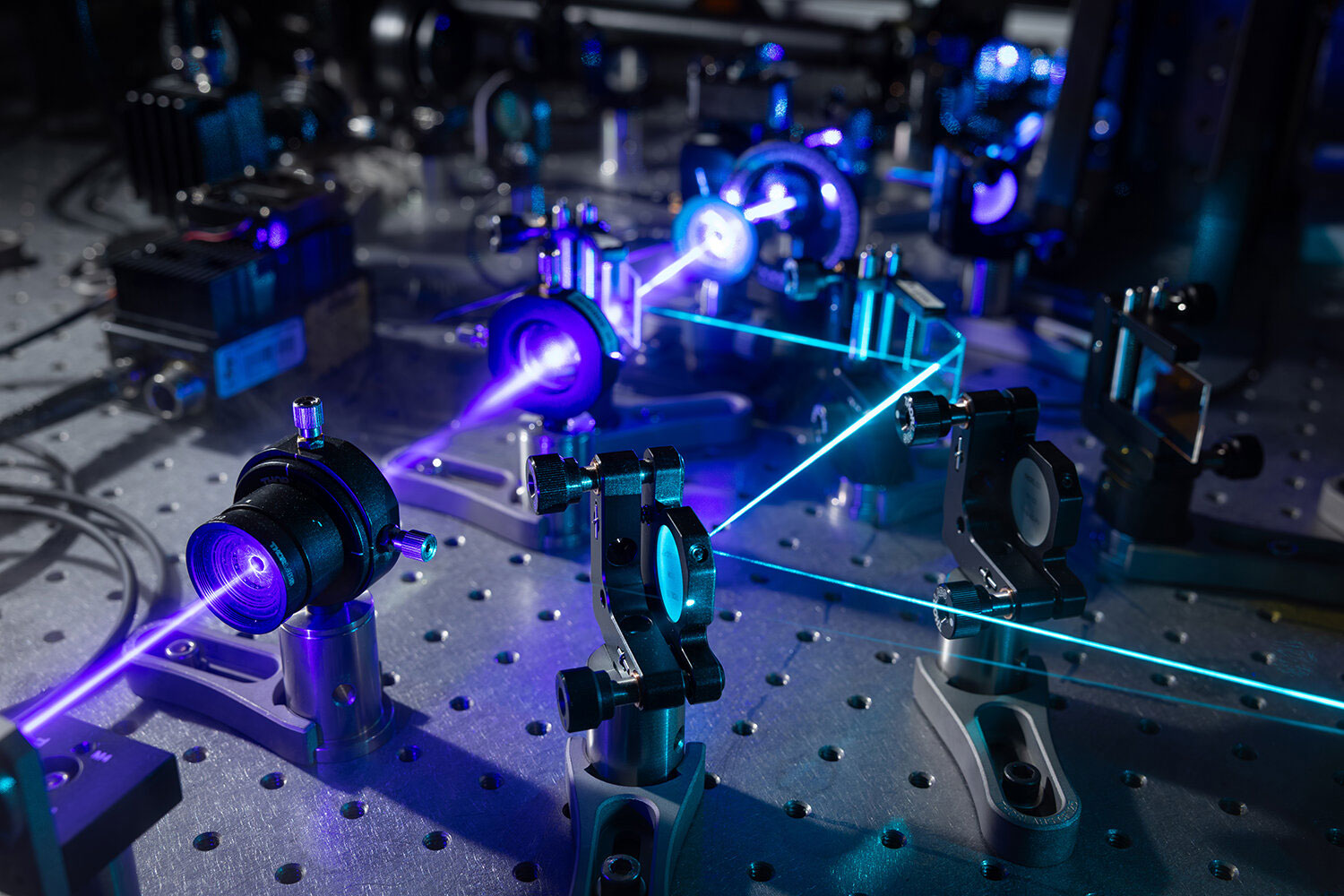
Stanford researchers have developed a microscope that can show how nanostructures interact inside living cells at the highest resolution achieved so far.
The view into living cells just got better.
Stanford researchers have merged two microscopy methods to build a unique instrument that can capture cell structures interacting in real time at an unprecedented resolution of 120 nanometers. It is the highest resolution yet achieved without fluorescent labels.
The technology, known as Interferometric Image Scanning Microscopy, or iISM, gives scientists a way to watch cellular structures in their broader environment, including how they react to invaders such as pathogens or to drugs. The advance is described in the journal Light: Science and Applications.
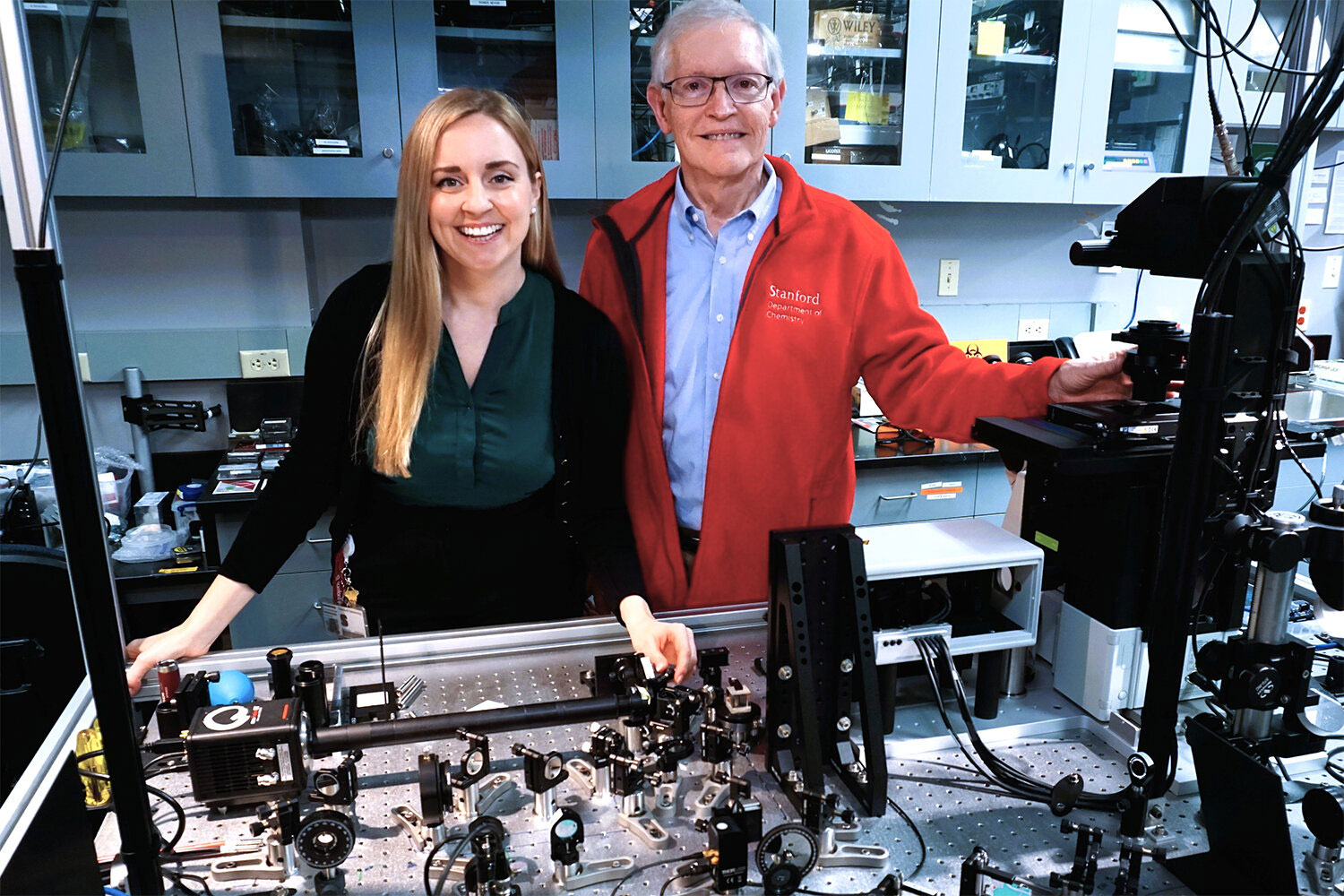
“This new microscope provides a fantastic new view into the cell, where you can see the tiny structures and machines in the cell moving, changing, and interacting without having to add fluorescence to observe them,” said senior author W.E. Moerner, the Harry S. Mosher Professor of Chemistry in Stanford’s School of Humanities and Sciences. “It’s a wonderful look into these complex little cellular boxes that drive our life.”
The abilities of iISM could support new discoveries across many areas of the life sciences, including research on disease mechanisms, drug development, and interactions between plants and microbes.
Although iISM does not reach the same resolution as some highly specialized microscopes, its label-free approach offers major benefits. Scientists can observe many cellular structures at the same time and follow them for longer periods. By comparison, fluorescence-based methods usually mark only a few selected structures at once. Fluorescent signals can also fade over time. In addition, the labels can be difficult to introduce and may sometimes alter the behavior of the structures being studied.
The iISM also works with much lower illumination power than similar high contrast label-free methods. That reduces the chance of light-related damage in living cells and makes it less likely that the imaging process will disturb the small, fragile structures under observation.
First author Michelle Kueppers, a postdoctoral scholar in Moerner’s lab, said the new microscope is not meant to replace fluorescence microscopy, which has produced important insights in biology for decades.
“Every method has its advantages and disadvantages, and we believe in a complementary implementation in the future,” Kueppers said. “If we use the strengths of fluorescence for molecular specificity and the strength of iISM for label-free context and dynamics, we can really start tackling questions that have been difficult to address before.”
Many ‘eyes’ on the same point
The iISM reaches higher resolution and sensitivity by combining the strengths of two microscopy approaches. That combination reflects the expertise of the two coauthors. Moerner, who received the 2014 Nobel Prize in chemistry for his work on super-resolution fluorescence microscopy, recruited Kueppers to Stanford because her doctoral research focused on “interferometric scattering microscopy.”
Scattering is the reason the sky appears blue. When light strikes small particles, as sunlight does when it passes through the atmosphere and encounters dust, water droplets, and other molecules, it changes direction and scatters. Particles in Earth’s atmosphere scatter short blue wavelengths more strongly than red wavelengths, making the sky look blue to human eyes.
In an interferometric scattering microscope, a laser shines on a cell, and tiny structures inside the cell scatter some of that light. A second laser beam boosts the faint scattered light enough for detection, allowing small structures to be seen.
The central advance in iISM comes from pairing interferometric scattering with an adapted idea from next-generation confocal microscopes. Traditional confocal microscopes use a pinhole and a single detector to focus on target structures. More advanced versions use camera-based array detectors that capture many views of the same region.
For iISM, the Stanford team used an array detector that collects more light than a pinhole and single detector system. This improves depth and precision. The concept is similar to how two human eyes gather information to separate foreground from background, except iISM uses tens to hundreds of views from an array detector rather than just two “eyes.” The researchers then created a method for combining those measurements into images with sharper detail and stronger contrast.
The result is a label-free microscope that can achieve about 120-nanometer resolution while using less laser power and preserving imaging speed. That means scientists can observe living cells for longer periods and with a gentler approach.
Wide vision for wide applications
Moerner and Kueppers are now working to improve the technology further and make it available to more scientists.
They have already begun three collaborations with other Stanford researchers. One project uses the microscope to watch interactions among plant cells, fungi, and bacteria in real time. Another uses iISM to observe how a cancer drug enters a cell. A third planned project will examine how red blood cells change shape when they encounter a malaria infection.
“This is not a niche technique,” Kueppers said. “It has broad applications, and we hope the life science community will be well served by it, leading to many new discoveries.”
Reference: “Interferometric Image Scanning Microscopy for label-free imaging at 120 nm lateral resolution inside live cells” by Michelle Küppers, and W. E. Moerner, 27 February 2026, Light: Science & Applications.
DOI: 10.1038/s41377-026-02210-y
This research received support from the U.S. National Institute of General Medical Sciences.
Never miss a breakthrough: Join the SciTechDaily newsletter.
Follow us on Google and Google News.
7 Comments
how two wok thi microphone wif laser
this is NOT me Jett Cook
hi guys
bitch
im kinda bald
i can see
i can fly