A new study sheds light on why promising cancer treatments can produce dramatically different results across patients.
One of the biggest challenges in cancer treatment is that a therapy that works well for one patient may completely fail in another. A study published in Nature Communications by a multidisciplinary team led by Dr. Louise Fets at the MRC Laboratory of Medical Sciences (LMS) examined how a class of targeted drugs called PARP inhibitors spreads through ovarian tumor samples.
Using advanced imaging techniques, the researchers found that these drugs can build up inside lysosomes, which are small structures within cells that act as “recycling centers.” This buildup allows the drugs to be stored and released over time, influencing how effective the treatment is.
Mapping drug delivery
Cancer treatments have advanced rapidly in recent years, leading to better outcomes for many patients. For ovarian cancer, PARP inhibitors have played a major role in this progress. However, not all patients benefit equally, and some develop resistance to these drugs.
For a treatment to work, the drug must accumulate inside cancer cells at levels high enough to kill them. Despite its importance, scientists still do not fully understand how drugs are distributed within tumors or what controls this process.
This study highlights that the issue is not only whether a drug reaches a tumor, but also how it spreads throughout the tumor and inside individual cells. The researchers used thin slices of ovarian tumors taken from patients and kept them alive in the lab. These samples, known as “explants,” were treated with PARP inhibitors so scientists could directly observe how the drugs moved through real human tumor tissue.
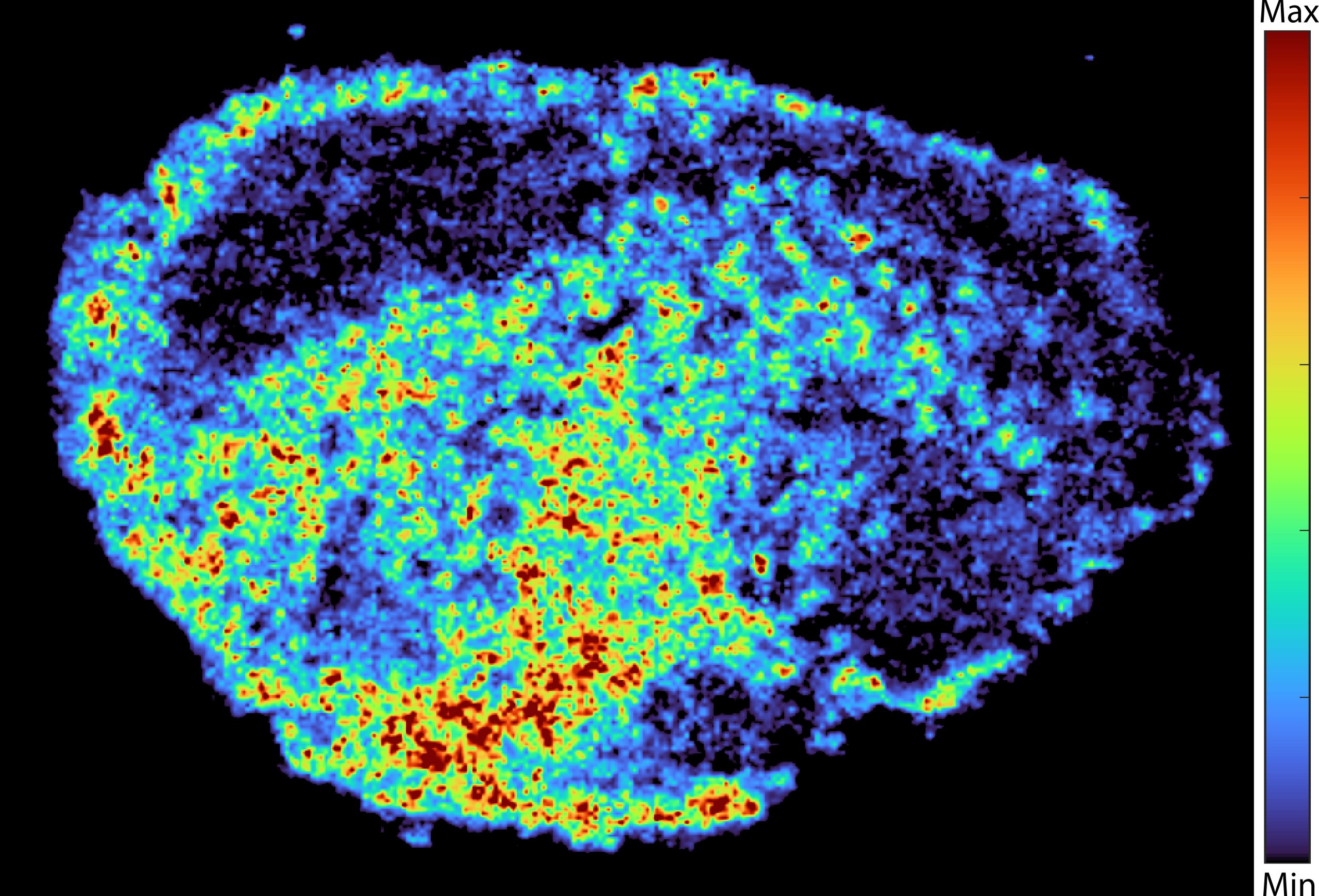
The team used mass spectrometry imaging to produce detailed maps showing where drug molecules collected. They combined this with spatial transcriptomics, which allowed them to compare gene activity in areas with high and low drug concentrations within the same sample. The findings showed large differences in drug levels across different regions of the same tumor and between patients, even when the same dose was used.
“A novel aspect of this study was the use of mass spectrometry imaging to directly measure and visualize drug uptake in patient tumor tissue. Through the spatial mapping of drug molecules, we could pinpoint regions of high and low drug and compare gene expression, from the same tissue slice, using spatial transcriptomics,” says Dr. Zoe Hall, senior author and Associate Professor at Imperial’s Department of Metabolism, Digestion and Reproduction.
Lysosomes: the cell’s hidden drug storehouses
The researchers found that lysosomes play a key role in this uneven distribution. Some PARP inhibitors are drawn into these compartments and stored there instead of spreading evenly throughout the cell. As a result, the drugs become trapped and form internal reservoirs.
These reservoirs act as slow-release storage sites, holding the drug and releasing it gradually over time. This leads to higher exposure in some cells, while others receive much less of the drug. Not all PARP inhibitors behave the same way. The study showed that drugs such as rucaparib and niraparib are affected by this process, while others like olaparib are not.
“We were surprised to see large variability in drug accumulation at the single-cell level. This variability was driven by the build-up of a drug in lysosomes, which are acting as reservoirs, increasing the exposure of cancer cells to drugs, by storing and releasing the drug when needed,” says Dr. Carmen Ramirez Moncayo, first author and Postdoctoral Researcher at the LMS.
The future of cancer treatment
PARP inhibitors are already widely used to treat ovarian, breast, and prostate cancers, and they are being tested in clinical trials for many other types of cancer. A better understanding of how these drugs are stored and distributed inside cells could lead to more personalized treatment strategies that improve outcomes and reduce the risk of resistance or relapse.
“By understanding how drugs are taken up into cells, we can understand whether this influences why cancer drugs work for some people and not for others. Eventually, we hope to be able to study the molecular signature of a patient’s tumor to help tailor therapeutic approaches in a more personalized way,” says Dr. Louise Fets, senior author and Head of the LMS’ Drug Transport and Tumor Metabolism Group.
This study used patient tumor tissue maintained outside the body. In actual patients, drugs are delivered through the bloodstream, where tumor blood vessels are often disorganized, which may further increase uneven drug distribution. Future research will use animal models and larger patient studies to better understand how drug delivery, tumor structure, and lysosomal storage interact in real clinical settings, including in relapsed cancers.
Reference: “Multimodal imaging reveals a lysosomal drug reservoir that drives heterogeneous distribution of PARP inhibitors” by Carmen R. Moncayo, Restuadi Restuadi, Guanying Zhang, Daniel Marks, Paula Ortega-Prieto, Emily Doherty, Nathalie Lambie, Chad Whilding, Ivan Andrew, Alex Montoya, Bhavik Patel, Katie Tyson, Betheney R. Pennycook, Lauren Pendergast, Vincen Wu, Zoltan Takats, Nik Matthews, George R. Young, Priyanka Verma, Pavel Shliaha, Laurence Game, Boris Lenhard, Iain McNeish, Christina Fotopoulou, Alexis R. Barr, Paula Cunnea, Zoe Hall and Louise Fets, 17 March 2026, Nature Communications.
DOI: 10.1038/s41467-026-70558-1
This research was supported by funding from the Medical Research Council, Cancer Research UK, a PhD studentship from the Integrative Toxicology Training Partnership administered by the MRC Toxicology Unit, and a Victoria’s Secret Global Fund for Women’s Cancers Career Development Award, in partnership with Pelotonia and AACR.
Never miss a breakthrough: Join the SciTechDaily newsletter.
Follow us on Google and Google News.
1 Comment
Thanks for sharing this knowledge.