
Researchers from the Max Born Institute in Berlin have successfully performed X-ray Magnetic Circular Dichroism (XMCD) experiments in a laser laboratory for the first time.
Unlocking the secrets of magnetic materials requires the right illumination. Magnetic x-ray circular dichroism makes it possible to decode magnetic order in nanostructures and to assign it to different layers or chemical elements. Researchers at the Max Born Institute in Berlin have succeeded in implementing this unique measurement technique in the soft-x-ray range in a laser laboratory. With this development, many technologically relevant questions can now be investigated outside of scientific large-scale facilities for the first time.
Magnetic nanostructures have long been part of our everyday life, e.g., in the form of fast and compact data storage devices or highly sensitive sensors. A major contribution to the understanding of many of the relevant magnetic effects and functionalities is made by a special measurement method: X-ray Magnetic Circular Dichroism (XMCD).
This impressive term describes a fundamental effect of the interaction between light and matter: In a ferromagnetic material, there is an imbalance of electrons with a certain angular momentum, the spin. If one shines circularly polarized light, which also has a defined angular momentum, through a ferromagnet, a clear difference in transmission for a parallel or anti-parallel alignment of the two angular momenta is observable — a so-called dichroism.
This circular dichroism of magnetic origin is particularly pronounced in the soft-x-ray region (200 to 2000 eV energy of the light particles, corresponding to a wavelength of only 6 to 0.6 nm), when considering the element-specific absorption edges of transition metals, such as iron, nickel, or cobalt, as well as rare earths, such as dysprosium or gadolinium. These elements are particularly important for the technical application of magnetic effects.
Ultrafast Magnetization Processes with Soft X-Rays
The XMCD effect allows for precisely determining the magnetic moment of the respective elements, even in buried layers in a material and without damaging the sample system. If the circularly polarized soft-x-ray radiation comes in very short femto- to picosecond (ps) pulses, even ultrafast magnetization processes can be monitored on the relevant time scale. Until now, access to the required x-ray radiation has only been possible at scientific large-scale facilities, such as synchrotron-radiation sources or free-electron lasers (FELs), and has thus been strongly limited.
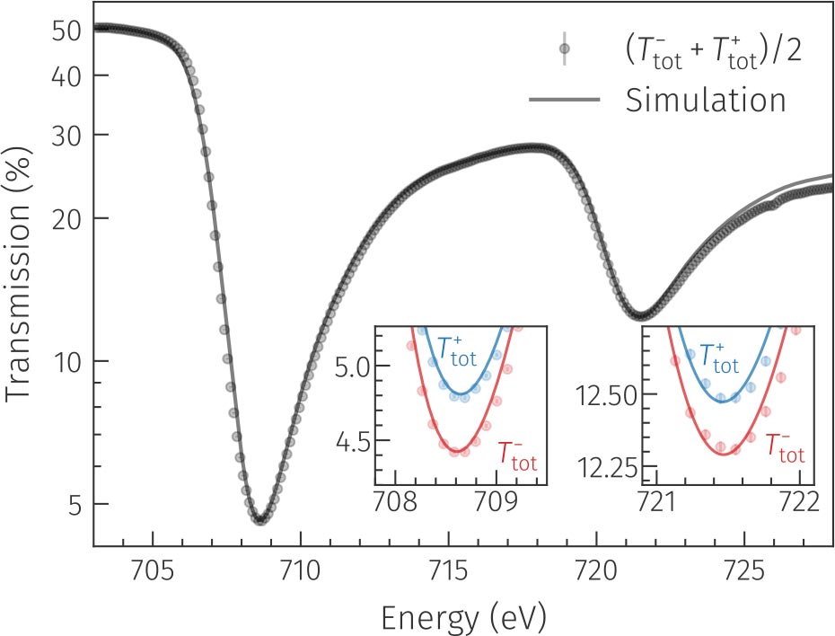
A team of researchers around junior research group leader Daniel Schick at the Max Born Institute (MBI) in Berlin has now succeeded for the first time in realizing XMCD experiments at the absorption L edges of iron at a photon energy of around 700 eV in a laser laboratory.
Laser-Driven Plasma
A laser-driven plasma source was used to generate the required soft x-ray light, by focusing very short (2 ps) and intense (200 mJ per pulse) optical laser pulses onto a cylinder of tungsten. The generated plasma thereby emits a lot of light continuously in the relevant spectral range of 200-2000 eV at a pulse duration of smaller than 10 ps. However, due to the stochastic generation process in the plasma, a very important requirement to observe XMCD is not met — the polarization of the soft-x-ray light is not circular, as required, but completely random, similar to that of a light bulb.
Therefore, the researchers used a trick: the x-ray light first passes through a magnetic polarization filter in which the same XMCD effect as described above is active. Due to the polarization-dependent dichroic transmission, an imbalance of light particles with parallel vs. anti-parallel angular momentum relative to the magnetization of the filter can be generated. After passing through the polarization filter, the partially circularly or elliptically polarized soft-x-ray light can be employed for the actual XMCD experiment on a magnetic sample.
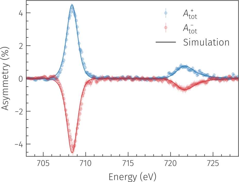
The work, published in the scientific journal OPTICA, demonstrates that laser-based x-ray sources are catching up with large-scale facilities. “Our concept for generating circularly polarized soft x-rays is not only very flexible but also partly superior to conventional methods in XMCD spectroscopy due to the broadband nature of our light source,” says the first author of the study and PhD student at the MBI, Martin Borchert. In particular, the already demonstrated pulse duration of the generated x-ray pulses of only a few picoseconds opens up new possibilities to observe and ultimately understand even very fast magnetization processes, e.g., when triggered by ultrashort light flashes.
Reference: “X-ray magnetic circular dichroism spectroscopy at the Fe L edges with a picosecond laser-driven plasma source” by Martin Borchert, Dieter Engel, Clemens von Korff Schmising, Bastian Pfau, Stefan Eisebitt and Daniel Schick, 4 April 2023, Optica.
DOI: 10.1364/OPTICA.480221
Never miss a breakthrough: Join the SciTechDaily newsletter.
Follow us on Google and Google News.