A new ultra-sensitive imaging system can make cancer cells light up, paving the way for faster and earlier detection.
Researchers have created a compact Raman imaging system that can reliably tell tumor tissue apart from normal tissue. The goal is to support earlier cancer detection and make molecular imaging easier to use beyond specialized research labs.
How SERS nanoparticles help tumors stand out
The system is built to pick up extremely faint signals from surface-enhanced Raman scattering (SERS) nanoparticles designed to attach to tumor markers. After these nanoparticles are placed on a sample or applied to the area being examined, the device reads their Raman signal and automatically flags locations that are more likely to contain tumor tissue.
“Traditional methods for cancer-related diagnosis are time-consuming and labor-intensive because they require staining tissue samples and having a pathologist look for any abnormalities,” said research team leader Zhen Qiu from the Institute for Quantitative Health Science and Engineering (IQ), Michigan State University. “While our system would not immediately replace pathology, it could serve as a rapid screening tool to accelerate diagnosis.”
Published results and stronger sensitivity than commercial systems
In Optica, Optica Publishing Group’s journal for high-impact research, Qiu and colleagues report that the system can separate cancerous cells from healthy cells while detecting Raman signals about four times weaker than a comparable commercial system. That jump in sensitivity comes from combining a swept-source laser — which changes wavelength during analysis — with a highly sensitive detector called a superconducting nanowire single-photon detector (SNSPD).
“This technology could eventually enable portable or intraoperative devices that enable clinicians to detect cancers at earlier stages, improve the accuracy of biopsy sampling, and monitor disease progression through less invasive testing,” said Qiu. “Ultimately, such advances could enhance patient outcomes and reduce diagnostic delays, accelerating the path from detection to treatment.”
Pushing detection limits with superconducting photon detection
Qiu’s lab investigates ways to use SNSPDs to upgrade different imaging platforms. SNSPDs rely on a superconducting wire that can register individual particles of light, allowing the system to capture extremely weak optical signals quickly, with very low noise.
For this work, the team set out to use an SNSPD to detect Raman signals far below what typical Raman systems can measure. Raman imaging reveals a sample’s chemical makeup by reading the distinct light-scattering fingerprints produced by its molecules. Those fingerprints can be boosted by using SERS nanoparticles.
“Combining this advanced detector with a swept-source Raman architecture that replaces a bulky camera and collects light more efficiently resulted in a system with a detection limit well beyond that of comparable commercial systems,” said Qiu. “Also, the fiber coupling configuration and compact design facilitate system miniaturization and future clinical translation.”
Testing tumor targeting and tissue contrast across samples
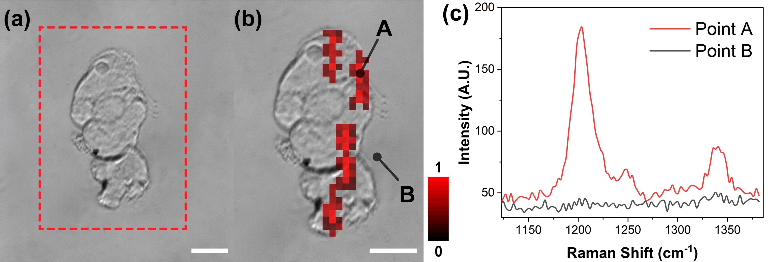
To evaluate performance, the researchers used SERS nanoparticles coated with hyaluronan acid, which helps the particles bind to CD44, a surface protein found on many tumor cells. They first tested simple solutions containing the nanoparticles and found the system could reach femtomolar sensitivity. Next, they used the platform on cultured breast cancer cells, mouse tumors, and healthy tissues.
“The SERS signals were strongly concentrated in tumor samples, with only minimal background detected in healthy tissue,” said Qiu. “This demonstrates both the system’s exceptional sensitivity and its ability to provide reliable tumor-versus-healthy contrast. Moreover, by adjusting or substituting the targeting molecule, this method could be adapted for other cancer types.”
What still needs to happen before clinical use
The researchers say clinical translation will require faster readout and broader validation. To increase speed, they are exploring alternative laser sources, such as VCSELs, and also testing whether narrowing the sweep range can help. They also plan multiplexing experiments using different nanoparticles to target multiple biomarkers simultaneously.
Reference: “High-sensitivity SNSPD-enabled swept source Raman spectroscopy for cancer detection with SERS nanoparticles” by Stephanie Boyd, Zhen Qiu, Gary D. Luker, Ming Han, Timothy M. Rambo, Aniwat Juhong, Cheng-you Yao, Jeremy S. Doredla, Aaron J. Miller, Xuefei Huang, Min Li, Yifan Liu, A. K. M. Atique Ullah and Bo Li, 19 December 2025, Optica.
DOI: 10.1364/OPTICA.569117
The researchers acknowledge industry collaborator Quantum Opus, which provided the SNSPD devices used in this work.
Never miss a breakthrough: Join the SciTechDaily newsletter.
Follow us on Google and Google News.