How the “Marsupial Sabertooth” Thylacosmilus Saw Its World
Study describes how extinct hypercarnivore likely achieved 3D vision despite wide-set eyes more characteristic of an herbivore than a predator.
A new study investigates how an extinct, carnivorous marsupial relative with canines so large they extended across the top of its skull could hunt effectively despite having wide-set eyes, like a cow or a horse. The skulls of carnivores typically have forward-facing eye sockets, or orbits, which helps enable stereoscopic (3D) vision, a useful adaptation for judging the position of prey before pouncing. Scientists from the American Museum of Natural History and the Instituto Argentino de Nivología, Glaciología, y Ciencias Ambientales in Mendoza, Argentina, studied whether the “marsupial sabertooth” Thylacosmilus atrox could see in 3D at all. Their results are published today (March 21) in the journal Communications Biology.
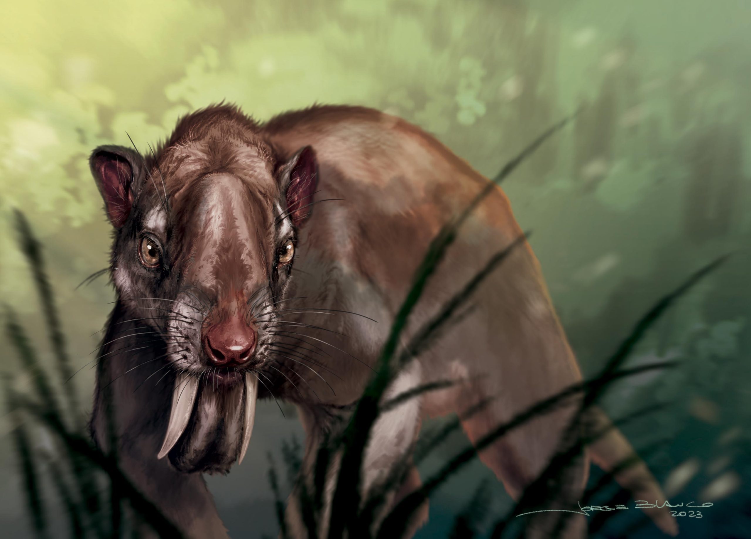
Popularly known as the “marsupial (or metatherian) sabertooth” because its extraordinarily large upper canines recall those of the more famous placental sabertooth that evolved in North America, Thylacosmilus lived in South America until its extinction about 3 million years ago. It was a member of Sparassodonta, a group of highly carnivorous mammals related to living marsupials.
Although sparassodont species differed considerably in size—Thylacosmilus may have weighed as much as 100 kilograms (220 pounds)—the great majority resembled placental carnivores like cats and dogs in having forward-facing eyes and, presumably, full 3D vision. By contrast, the orbits of Thylacosmilus, a supposed hypercarnivore—an animal with a diet estimated to consist of at least 70 percent meat—were positioned like those of an ungulate, with orbits that face mostly laterally. In this situation, the visual fields do not overlap sufficiently for the brain to integrate them in 3D.
Why would a hypercarnivore evolve such a peculiar adaptation? A team of researchers from Argentina and the United States set out to look for an explanation.
Canine Growth and Cranial Reorganization
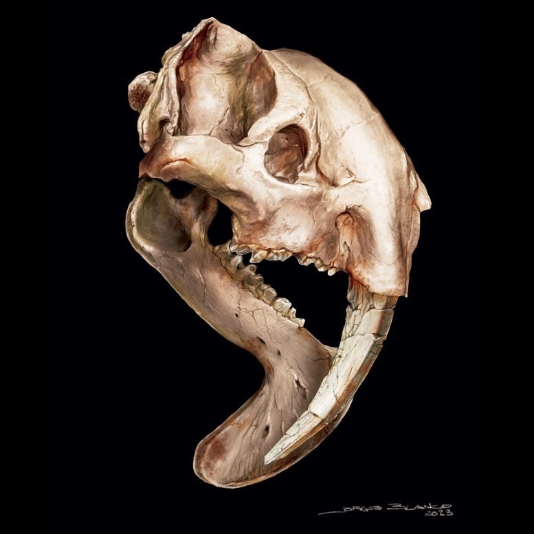
“You can’t understand cranial organization in Thylacosmilus without first confronting those enormous canines,” said lead author Charlène Gaillard, a Ph.D. student in the Instituto Argentino de Nivología, Glaciología, y Ciencias Ambientales (INAGLIA). “They weren’t just large; they were ever-growing, to such an extent that the roots of the canines continued over the tops of their skulls. This had consequences, one of which was that no room was available for the orbits in the usual carnivore position on the front of the face.”
Gaillard used CT scanning and 3D virtual reconstructions to assess orbital organization in a number of fossil and modern mammals. She was able to determine how the visual system of Thylacosmilus would have compared to those in other carnivores or other mammals in general. Although low orbital convergence occurs in some modern carnivores, Thylacosmilus was extreme in this regard: it had an orbital convergence value as low as 35 degrees, compared to that of a typical predator, like a cat, at around 65 degrees.
However, good stereoscopic vision also relies on the degree of frontation, which is a measure of how the eyeballs are situated within the orbits. “Thylacosmilus was able to compensate for having its eyes on the side of its head by sticking its orbits out somewhat and orienting them almost vertically, to increase visual field overlap as much as possible,” said co-author Analia M. Forasiepi, also in INAGLIA and a researcher in CONICET, the Argentinian science and research agency. “Even though its orbits were not favorably positioned for 3D vision, it could achieve about 70 percent of visual field overlap—evidently, enough to make it a successful active predator.”
Morphological Trade-offs for Functionality
“Compensation appears to be the key to understanding how the skull of Thylacosmilus was put together,” said study co-author Ross D. E. MacPhee, a senior curator at the American Museum of Natural History. “In effect, the growth pattern of the canines during early cranial development would have displaced the orbits away from the front of the face, producing the result we see in adult skulls. The odd orientation of the orbits in Thylacosmilus actually represents a morphological compromise between the primary function of the cranium, which is to hold and protect the brain and sense organs, and a collateral function unique to this species, which was to provide enough room for the development of the enormous canines.”
Lateral displacement of the orbits was not the only cranial modification that Thylacosmilus developed to accommodate its canines while retaining other functions. Placing the eyes on the side of the skull brings them close to the temporal chewing muscles, which might result in deformation during eating. To control for this, some mammals, including primates, have developed a bony structure that closes off the eye sockets from the side. Thylacosmilus did the same thing—another example of convergence among unrelated species.
The Mystery of Ever-Growing Canines
This leaves a final question: What purpose would have been served by developing huge, ever-growing teeth that required re-engineering of the whole skull?
“It might have made predation easier in some unknown way,” said Gaillard, “But, if so, why didn’t any other sparassodont—or for that matter, any other mammalian carnivore—develop the same adaptation convergently? The canines of Thylacosmilus did not wear down, like the incisors of rodents. Instead, they just seem to have continued growing at the root, eventually extending almost to the rear of the skull.”
Forasiepi underlined this point, saying, “To look for clear-cut adaptive explanations in evolutionary biology is fun but largely futile. One thing is clear: Thylacosmilus was not a freak of nature, but in its time and place it managed, apparently quite admirably, to survive as an ambush predator. We may view it as an anomaly because it doesn’t fit within our preconceived categories of what a proper mammalian carnivore should look like, but evolution makes its own rules.”
Reference: “Seeing through the eyes of the sabertooth Thylacosmilus atrox (Metatheria, Sparassodonta” by Charlène Gaillard, Ross D. E. MacPhee and Analía M. Forasiepi, 21 March 2023, Communications Biology.
DOI: 10.1038/s42003-023-04624-5
Never miss a breakthrough: Join the SciTechDaily newsletter.
Follow us on Google and Google News.
2 Comments
Seems when it opens its mouth there is some overlap opportunity for each eye. However, it could just be that each eye scans quickly enough to perceive in 3D, and also, if the eyes have no overlap or very little overlap, it’s also possible for the brain to make some assumptions that correlate with objective reality enough of the time to have a 3D understanding of the environment. For example, any amount of movement perceivable to the brain through the eyes could indicate information about 3D inferences as pixels move from one eye to the other. The apex where we would expect the overlap to be, but isn’t, still presents a wealth of information, even if we don’t understand fully how the information was used.
Basically, the animal was designed to eventually bite it’s own head off.