UW–Madison researchers have created a non-destructive method to study nacre layer thickness using hyperspectral interference tomography, revealing biological and paleoclimate insights.
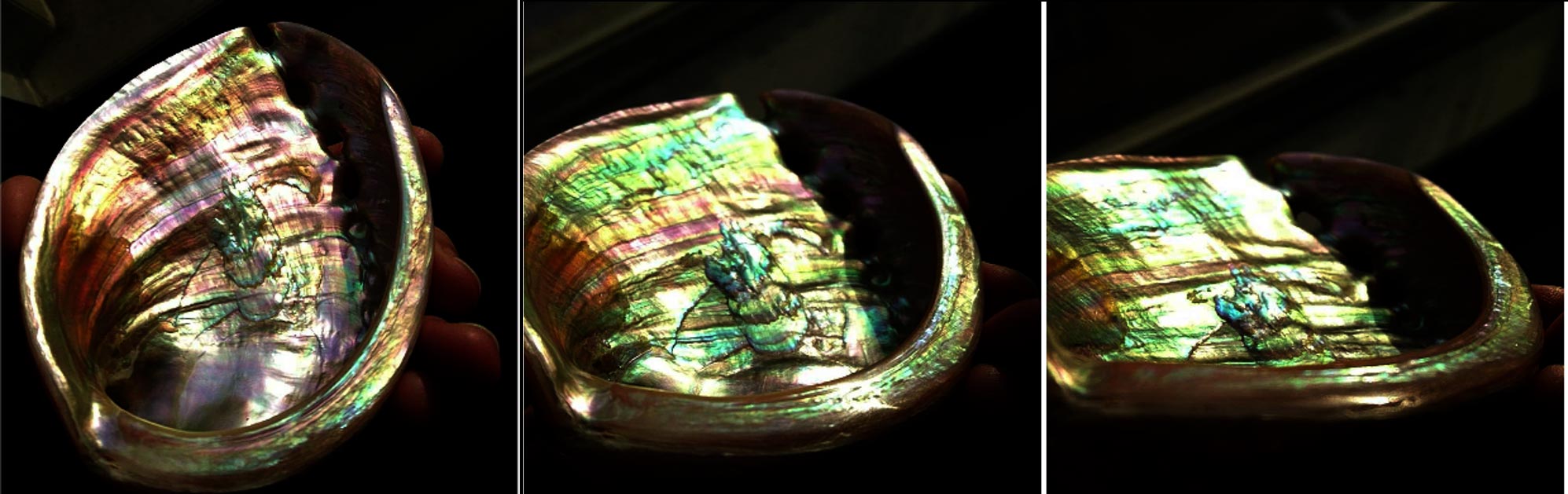
Most people know mother-of-pearl, an iridescent biomineral also called nacre, from buttons, jewelry, instrument inlays, and other decorative flourishes. Scientists, too, have admired and marveled at nacre for decades, not only for its beauty and optical properties but because of its exceptional toughness.
“It’s one of the most-studied natural biominerals,” says Pupa Gilbert, a University of Wisconsin–Madison physics professor who has studied nacre for more than a decade. “It may not look like much — just a shiny, decorative material. But it can be 3,000 times more resistant to fracture than aragonite, the mineral from which it’s made. It has piqued the interest of materials scientists because making materials better than the sum of their parts is extremely attractive.”
Now, a new, nondestructive optical technique will unlock even more knowledge about nacre, and in the process could lead to a new understanding of climate history. Gilbert, UW–Madison electrical engineering professor Mikhail Kats, their students and collaborators described the technique, called hyperspectral interference tomography, today in the journal Proceedings of the National Academy of Sciences.
Gilbert has learned how nacre forms, orders, resists fracture, and how its layered structure records the temperature at which it formed. This layered structure of nacre reflects light and generates different colors depending on layer thickness. That led to an interest in finding a way to assess the thickness of the nacre layers that doesn’t involve destroying the mollusk shell in which it is deposited.
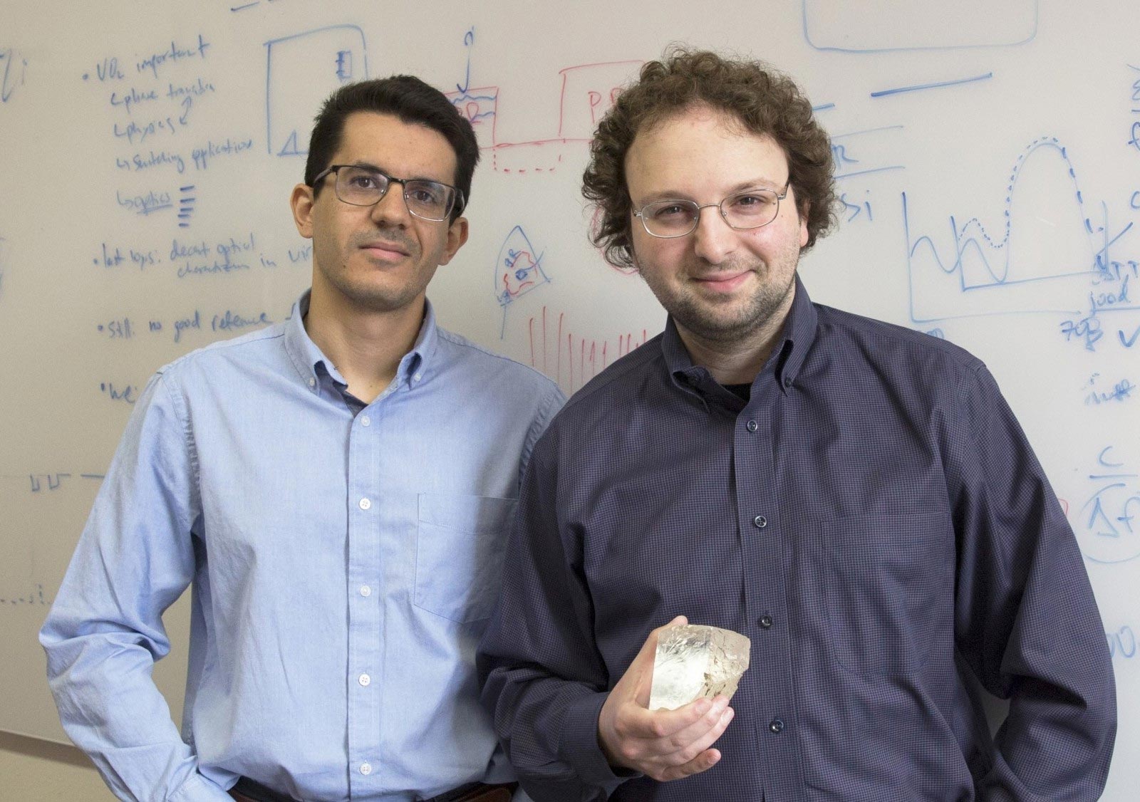
For help tackling that challenge, Gilbert turned to Kats and graduate student Jad Salman, who are experts in studying optical phenomena.
Imaging the Unimaginable
For the project, Salman prepared 22 fresh red abalone shells for optical analysis. But taking optical spectra of nacre is more difficult than it may seem.
“If you want to probe this type of shell, which has a curved topography, it’s very difficult to get a good spectrum with a conventional spectrometer,” Salman says.
That’s why the team turned to a newer technology, hyperspectral photography, to image the entire spectrum of the shell. Early on, they imaged the shells at industry partner Middleton Spectral Vision before acquiring their own hyperspectral camera.

Hyperspectral Interference Tomography
“It’s an imaging spectrometer where each pixel in the image gives a full spectrum,” says Salman. “When we use the camera in our setup, we can easily extract reliable spectral data over the large, uneven surface of a shell in one shot.”
In addition to the red abalone, the team also imaged the nacre of another species, paua shell from New Zealand, also called rainbow abalone. Salman then used sophisticated modeling software he developed to determine the thickness of the nacre layers pixel by pixel using the hyperspectral data.
The team is calling the combination of techniques hyperspectral interference tomography and anticipates that it will be applicable to measuring other transparent, layered structures found in plants, animals, geological samples or synthetic materials.
For Gilbert, the new technique revealed a surprise about red abalone; it showed for the first time that the thickness of nacre layers thins as the mollusk ages. Because this thickness records the temperature of the sea water in which it forms, the team believes it may be possible to use the technique to analyze fossil mollusk shells to learn about past climates.
“This project is made of a few different parts, each of those somewhat well understood,” says Kats. “The power of this research is that we brought all of this experimental and theoretical expertise, and were able to model not only engineered, well-behaved layered structures, but messy, disordered biological structures. And we were able to get useful information out of it in a way that a biologist or paleoclimatologist can use.”
Reference: “Hyperspectral interference tomography of nacre” by Jad Salman, Cayla A. Stifler, Alireza Shahsafi, Chang-Yu Sun, Stephen C. Weibel, Michel Frising, Bryan E. Rubio-Perez, Yuzhe Xiao, Christopher Draves, Raymond A. Wambold, Zhaoning Yu, Daniel C. Bradley, Gabor Kemeny, Pupa U. P. A. Gilbert and Mikhail A. Kats, 13 April 2021, Proceedings of the National Academy of Sciences.
DOI: 10.1073/pnas.2023623118
Other contributors to the research from UW–Madison include Cayla Stifler, Alireza Shahsafi, Chang-Yu Sun, Michel Frising, Bryan Rubio-Perez, Yuzhe Xiao, Raymond Wambold, Zhaoning Yu and Daniel Bradley. Stephen Weibel, Christopher Draves and Gabor Kemeny of Middleton Spectral Vision also contributed to the study.
This research was supported by grants from the Air Force Office of Scientific Research (FA9550-18-1-0146), Department of Energy (DE-FG02-07ER15899) and National Science Foundation (DMR-1603192).
Never miss a breakthrough: Join the SciTechDaily newsletter.
Follow us on Google and Google News.