
Molecular discoveries may help improve treatments for labor and pain.
When labor starts, the uterus must produce steady, precisely timed contractions to allow a safe delivery. Hormones such as progesterone and oxytocin are known to drive this process, but researchers have long believed that physical forces also matter. These forces include the stretching and pressure that build throughout pregnancy and intensify during birth.
A new study from Scripps Research, published in Science, explains how the uterus detects and responds to these forces at the molecular level. The findings offer new insight into why labor sometimes slows or begins too early and could guide future strategies to improve maternal care.
Pressure-sensing guides contractions
“As the fetus grows, the uterus expands dramatically, and those physical forces reach their peak during delivery,” says senior author Ardem Patapoutian, a Howard Hughes Medical Institute Investigator and the Presidential Endowed Chair in Neurobiology at Scripps Research. “Our study shows that the body relies on special pressure sensors to interpret these cues and translate them into coordinated muscle activity.”
Patapoutian shared the 2021 Nobel Prize in Physiology or Medicine for discovering the sensors that allow cells to sense touch and pressure. These sensors are ion channels made from the proteins PIEZO1 and PIEZO2, which enable cells to respond to mechanical force.
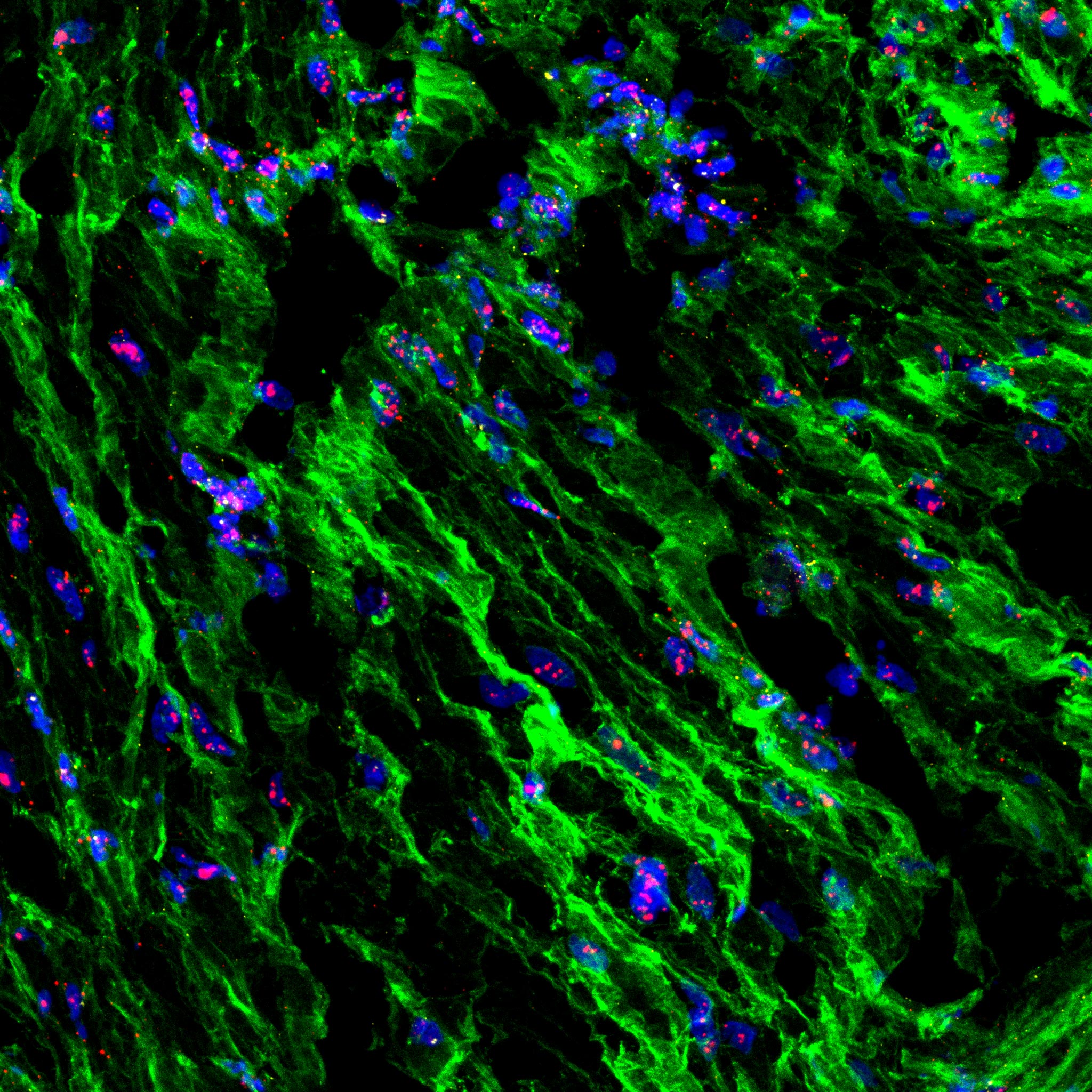
In the new research, Patapoutian and his colleagues found that PIEZO1 and PIEZO2 play different but complementary roles during childbirth. PIEZO1 is most active in the smooth muscle of the uterus, where it detects rising pressure as contractions strengthen.
PIEZO2 is located in sensory nerves in the cervix and vagina and is activated by stretching caused by the descending fetus, boosting uterine contractions through a neural reflex. Together, these proteins convert stretch and pressure into electrical and chemical signals that synchronize uterine contractions. Each pathway can partly support the other, helping labor continue even if one is weakened.
Mouse experiments reveal cooperation
To test how these pathways work, the researchers used mouse models in which PIEZO1 and PIEZO2 were selectively removed from either uterine muscle or the sensory nerves around the cervix and vagina. Sensors placed in pregnant mice measured contraction strength and timing during natural labor. Animals lacking both proteins showed weaker uterine pressure and delayed delivery, indicating that muscle-based and nerve based sensing act together and that losing both greatly disrupts labor.
Additional experiments showed that PIEZO signaling controls the production of connexin 43, a protein that forms gap junctions. These tiny channels connect neighboring smooth muscle cells so they contract as a unit. When PIEZO signaling was absent, connexin 43 levels fell, and the coordination between muscle cells broke down.
“Connexin 43 is the wiring that allows all the muscle cells to act together,” says first author Yunxiao Zhang, a postdoctoral research associate in Patapoutian’s lab. “When that connection weakens, contractions lose strength.”
Human tissue supports the model
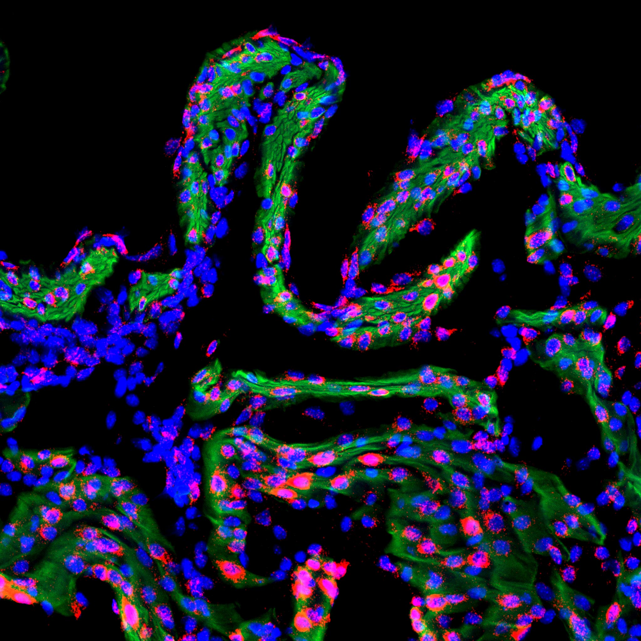
Additionally, human uterine tissue samples showed similar PIEZO1 and PIEZO2 expression patterns as those in mice, suggesting that a comparable force-sensing mechanism may operate in people, too. This could help explain certain labor complications, such as weak or irregular contractions that prolong delivery. Together, the findings are consistent with clinical observations that complete sensory nerve block causes prolonged labor during childbirth.
“In clinical practice, epidurals are given in carefully controlled doses because blocking sensory nerves completely can make labor much longer,” notes Zhang. “Our data mirror that phenomenon; when we removed the sensory PIEZO2 pathway, contractions weakened, suggesting that some nerve feedback promotes labor.”
Paths toward new therapies
The research team’s results open possibilities for more refined approaches to labor management and pain relief. If scientists can identify molecules that modulate PIEZO activity safely, they may one day be able to dampen or enhance uterine contractions as needed.
For mothers at risk of preterm labor, a PIEZO1 blocker—if developed—to slow contractions could complement existing drugs that relax muscle tissue by limiting calcium entry into cells. Conversely, a compound that activates PIEZO channels might help strengthen contractions in stalled labor.
Although such clinical applications remain distant, the foundational science continues to take shape. The research team is now investigating how PIEZO signaling interacts with hormonal pathways that regulate pregnancy.
Prior studies have shown that progesterone—the hormone that keeps the uterus relaxed during pregnancy—can suppress connexin 43 expression even when PIEZO channels are active, ensuring contractions don’t start prematurely. When progesterone levels drop near term, the PIEZO-driven calcium signals may help initiate the chain of biological events that lead to delivery.
“PIEZO channels and hormonal cues are two sides of the same system,” points out Zhang. “Hormones set the stage, and force sensors help determine when and how strongly the uterus contracts.”
Future work will delve deeper into the nerve pathways involved, since not all sensory fibers around the uterus contain PIEZO2. Some may respond to other stimuli and serve as backups when PIEZO2 is absent. Understanding which sensory nerves promote labor versus which convey pain could eventually lead to more precise forms of pain control that don’t slow delivery.
For now, the findings establish that the body’s ability to sense force isn’t limited to touch or balance—it’s also vital for one of life’s most fundamental biological events.
“Childbirth is a process where coordination and timing are everything,” says Patapoutian. “We’re now starting to understand how the uterus acts as both a muscle and a metronome to ensure that labor follows the body’s own rhythm.”
Reference: “PIEZO channels link mechanical forces to uterine contractions in parturition” by Yunxiao Zhang, Sejal A. Kini, Sassan A. Mishkanian, Oleg Yarishkin, Renhao Luo, Saba Heydari Seradj, Verina H. Leung, Yu Wang, M. Rocío Servín-Vences, William T. Keenan, Utku Sonmez, Manuel Sanchez-Alavez, Yuejia Liu, Xin Jin, Darren J. Lipomi, Li Ye, Michael Petrascheck, Antonina I. Frolova, Sarah K. England and Ardem Patapoutian, 13 November 2025, Science.
DOI: 10.1126/science.ady3045
This work was supported by the Abide-Vividion Foundations; the Baxter Foundation; the BRAIN Initiative; the Chan Zuckerberg Initiative; the Dana Foundation; the Dorris Scholar Award; the George E. Hewitt Foundation for Medical Research postdoctoral fellowship; the Howard Hughes Medical Institute Investigators; the Merck Fellow of the Damon Runyon Cancer Research Foundation (DRG-2405-20); the National Institutes of Health (NIH Director’s New Innovator Award DP2DK128800, and grants R35 NS105067, R01 AT012051 and R01 AG067331); the National Science Foundation (grant CMMI-2135428); the WashU Reproductive Specimen Processing and Banking Biorepository (ReProBank); and the Whitehall Foundation.
Never miss a breakthrough: Join the SciTechDaily newsletter.
Follow us on Google and Google News.
1 Comment
Childbirth women shouldn’t be laying down this causes non stop bleeding in Africa and Japan the women “Must” stand or sit on a sitz chair then the baby slides out C-Section cut top to bottom NOT left to right during pregnancy do a calm stretch muscle exercise. Play classical music for the baby to hear talk to the baby.