Researchers at NYU have mapped the brain’s auditory corollary discharge pathways, which help differentiate self-generated sounds from external noises, in epilepsy patients.
The study identifies the motor cortex’s role in managing these signals and provides insights into schizophrenia by explaining auditory hallucinations as disruptions in these pathways.
Understanding Auditory Corollary Discharge
Experiments involving people with epilepsy have revealed the brain’s electrical circuits that enable it to distinguish self-generated sounds, like one’s own voice, from background noise.
These signals, known as auditory corollary discharges, originate and terminate in specific regions of the brain’s folded outer layer, or cortex. The motor cortex, responsible for voluntary muscle movements, including those used in speech, works in tandem with the auditory cortex, which governs hearing.
Evolutionary Perspectives and Clinical Implications
From an evolutionary perspective, the ability to differentiate one’s own sounds from those of others has likely played a critical role in survival. This mechanism, powered by rapid, milliseconds-long electrical signals, helps animals like crickets discern their own mating chirps, allows songbirds to focus on their songs, and enables bats to navigate using sound echoes.
In humans, disruptions to this system are also thought to be hallmarks of auditory hallucinations, or “hearing voices,” in people with schizophrenia who cannot distinguish real voices from outside sounds, say the study authors. Disturbances in auditory corollary discharge signals are also thought to be involved in stuttering.
Challenges in Human Brain Signal Research
Previous experiments had tracked this electrical brain circuit to the motor cortex in other mammals, but the field has struggled to determine where discharge signals originate in the human motor cortex. This is partly because of the difficulty in recording brain activity while people are awake and talking, but mainly due to complexity of the computer analysis needed to analyze the recordings.
NYU Study on Auditory Signals in Epilepsy Patients
For the new study, led by researchers at NYU Langone Health, its Neuroscience Institute, and NYU Tandon School of Engineering, neuroscientists conducted voice experiments in eight adults with epilepsy. All were undergoing routine surgery to determine the source of their seizures and volunteered to participate in word exercises.
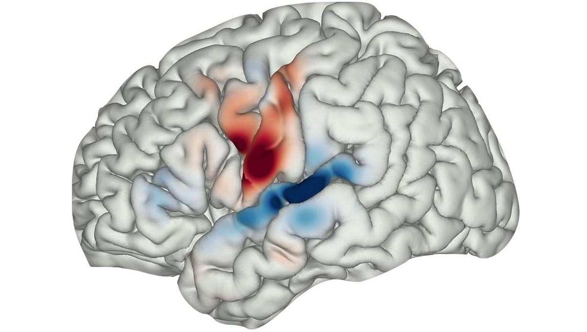
Published on December 3 in the Proceedings of the National Academy of Sciences (PNAS), the report describes how the researchers mapped auditory corollary discharge signals from the bottom, or ventral, part of the motor cortex, a subregion called the precentral gyrus. The electrical signals, lasting on average 120 milliseconds, were then found to move down and across the folds of the precentral gyrus to a neighboring auditory cortical subregion, called the superior temporal gyrus.
Key Findings and Insights into Schizophrenia
“We believe our study solves a long-standing puzzle in our understanding of human speech, offering the first direct evidence of the motor cortex brain circuits involved in corollary discharge that allow us to stay alert to our surroundings even while we are speaking,” said study lead investigator Amirhossein Khalilian-Gourtani, PhD. Dr. Khalilian-Gourtani is a postdoctoral research fellow in the Department of Neurology at NYU Grossman School of Medicine.
“Our findings also provide specific insight into schizophrenia, offering an explanation for the source of auditory hallucinations, as resulting from disrupted corollary discharge between the brain’s motor and auditory cortices,” said neuroscientist Adeen Flinker, PhD, a study senior investigator.
“What we and many other researchers suspect is happening in some people with schizophrenia is that they are unable to dissociate their own voice from others or even other external stimuli,” said Dr. Flinker, an associate professor in the Department of Neurology and NYU Tandon School of Engineering.
Methodology and Data Analysis of the Study
As part of the new study, researchers made more than 3,200 recordings of electrical brain activity while patients completed a series of voice experiments during planned breaks in their surgery. All patients had upward of 200 probes inserted into their brains to primarily monitor any seizure-related electrical activity. The research team then used a computer model to assess and predict what regions were active in the corollary discharge during speech in the word experiments designed to track the discharge.
In one of the exercises, patients were asked to listen to and then repeat a word, such as “balloon”; complete a sentence with the same word when answering the question “The boy blew up a … ?”; and look at a picture of a balloon and describe it with the same word.
Each test required the patient to tune out what word they were hearing while still being alert to their visual and acoustic surroundings, staying focused, and saying aloud the same word.
Study participants were mostly men and women in their 30s and 40s and had been recorded since 2019 at NYU Langone. Researchers recorded electrical activity inside most subregions of the patients’ brains as the patients heard themselves responding to recordings of statements being read aloud by others. Such audio-feedback tests have been developed to safely study how the human brain learns and processes speech.
Future Research Directions
Dr. Flinker says the team plans tests to assess further how and whether the corollary discharge circuit is active immediately before hallucinations induced during brain stimulation. They also have plans to work with psychiatrists on noninvasive means of testing the signal in people with schizophrenia.
Reference: “A corollary discharge circuit in human speech” by Amirhossein Khalilian-Gourtani, Ran Wang, Xupeng Chen, Leyao Yu, Patricia Dugan, Daniel Friedman, Werner Doyle, Orrin Devinsky, Yao Wang and Adeen Flinker, 3 December 2024, Proceedings of the National Academy of Sciences.
DOI: 10.1073/pnas.2404121121
Funding support for the study was provided by National Science Foundation grant IIS-1912286 and National Institutes of Health grants R01NS109367, R01NS115929, and R01DC018805.
Besides Dr. Khalilian-Gourtani and Dr. Flinker, other NYU Langone and NYU Tandon researchers involved in the study are co-investigators Ran Wang; Xufeng Chen; Leyao Yu; Patricia C. Dugan, MD; Daniel Friedman, MD; Werner Doyle; Orrin Devinsky, MD; and Yao Wang, PhD.
Never miss a breakthrough: Join the SciTechDaily newsletter.
Follow us on Google and Google News.