Duke researchers study an approach that may help millions manage diabetic neuropathy and chemotherapy-induced nerve pain.
For millions of people with chronic nerve pain, even gentle contact can trigger intense discomfort. Researchers have suspected that injured nerve cells struggle because their internal energy producers, known as mitochondria, stop working efficiently.
A study published in Nature now points to a potential solution: restoring healthy mitochondria to damaged nerve cells.
Rebuilding nerve cell energy
In experiments using human tissue and mouse models, scientists at Duke University School of Medicine found that replacing or boosting mitochondria significantly reduced pain associated with diabetic neuropathy and chemotherapy-induced nerve damage. In some cases, pain relief lasted as long as 48 hours.
Rather than simply dulling symptoms, this strategy aims to correct what researchers believe is an underlying cause of neuropathic pain: disrupted energy supply within nerve cells. When mitochondria fail, cells cannot generate the energy required to maintain normal function and resist inflammation.
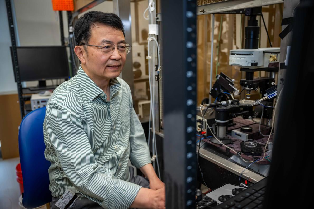
“By giving damaged nerves fresh mitochondria — or helping them make more of their own — we can reduce inflammation and support healing,” said the study’s senior author Ru-Rong Ji, PhD, director of the Center for Translational Pain Medicine in the Department of Anesthesiology at Duke School of Medicine. “This approach has the potential to ease pain in a completely new way.”
The findings add to growing evidence that cells can transfer mitochondria between one another. Scientists increasingly view this exchange as a natural support mechanism that may influence a range of conditions, including obesity, cancer, stroke, and chronic pain.
The secret life of glial cells
The researchers uncovered an unexpected role for satellite glial cells, which surround sensory neurons. These glial cells appear to pass mitochondria to neurons through tiny structures known as tunneling nanotubes.
When this transfer process breaks down, nerve fibers begin to deteriorate, leading to symptoms such as pain, tingling, and numbness, particularly in the hands and feet, where nerve endings are most distant from the spinal cord.
“By sharing energy reserves, satellite glial cells may help keep neurons out of pain,” said Ji, a professor of anesthesiology, neurobiology, and cell biology at Duke School of Medicine.
Enhancing this mitochondrial exchange reduced pain behaviors in mice by up to 50%, according to the study.
The team also tested a more direct method. They injected isolated mitochondria from either human or mouse sources into the dorsal root ganglia, a cluster of nerve cells that transmit sensory signals to the brain. This intervention produced similar pain relief, but only when the transplanted mitochondria were healthy. Mitochondria taken from individuals with diabetes did not produce the same benefit.
Identifying the molecular machinery
Further analysis identified a protein called MYO10 as crucial for forming the nanotubes that enable mitochondrial transfer between cells.
Ji conducted the research with lead author Jing Xu, PhD, a research scholar in the Department of Anesthesiology, and collaborator Caglu Eroglu, PhD, a Duke professor of cell biology known for her work on glial cells.
Additional studies are needed, including high-resolution imaging to observe precisely how nanotubes deliver mitochondria within living nerve tissue.
Still, the research reveals a previously underappreciated communication pathway between nerve cells and glial cells. By targeting cellular energy transfer, this approach could offer a new way to address chronic nerve pain at its biological source rather than simply suppressing symptoms.
Reference: “Mitochondrial transfer from glia to neurons protects against peripheral neuropathy” by Jing Xu, Yize Li, Charles Novak, Min Lee, Zihan Yan, Sangsu Bang, Aidan McGinnis, Sharat Chandra, Vivian Zhang, Wei He, Terry Lechler, Maria Pia Rodriguez Salazar, Cagla Eroglu, Matthew L. Becker, Dmitry Velmeshev, Richard E. Cheney and Ru-Rong Ji, 7 January 2026, Nature.
DOI: 10.1038/s41586-025-09896-x
National Institutes of Health, Department of Defense and Duke Department of Anesthesiology helped fund the study with authors receiving additional support from the Paul and Daisy Soros Fellowship, Howard Hughes Medical Institute, Michael J. Fox and the Aligning Science Across Parkinson’s Initiative, and Duke University Neurobiology Research Fund.
Never miss a breakthrough: Join the SciTechDaily newsletter.
Follow us on Google and Google News.
2 Comments
I can’t comment for the mice but as far as human subjects go it appears to be another case of “can’t see the forest for the trees.” In consideration of the symptoms they are aware of and want to treat, inflammation, failed mitochondria, obesity, cancer, stroke and chronic pain, it sounds to me like the sub-acute nearly subclinical non-IgE-mediated food (minimally) allergy reactions aggravated (or not) with toxic FDA approved food additives and excessive medical errors I’ve been commenting about online for years. And so it goes, because mainstream medicine failed to recognize Dr. Arthur F. Coca’s contributions ninety years ago, thousands of researchers keep independently duplicating each other’s mistakes. For a wise change of direction: https://odysee.com/@charlesgshaver:d?view=about
Well put articulated just right. Always going to big those factors that how much is the other notice perspective
. How we view the situation