A new imaging technology allows researchers to map human tissue development using organoids, aiding disease research and the creation of a comprehensive tissue atlas.
Which types of cells can be located in various human tissues, and where? Which genes show activity in these individual cells, and which proteins can be identified within them? Detailed answers to these inquiries and more are expected to be supplied by a specialized atlas. This atlas will particularly elucidate how different tissues take shape during embryonic development and the underlying causes of diseases.
In the process of developing this atlas, the researchers have the goal to chart not just tissues directly procured from humans but also structures referred to as organoids. These are three-dimensional tissue aggregates that are grown in the lab and develop in a manner similar to human organs, albeit on a smaller scale.
“The advantage of organoids is that we can intervene in their development and test active substances on them, which allows us to learn more about healthy tissue as well as diseases,” explains Barbara Treutlein, Professor of Quantitative Developmental Biology at the Department of Biosystems Science and Engineering at ETH Zurich in Basel.
To help produce such an atlas, Treutlein, together with researchers from the Universities of Zurich and Basel, has now developed an approach to gather and compile a great deal of information about organoids and their development. The research team applied this approach to the organoids of the human retina, which they derived from stem cells.
Many Proteins Visible Simultaneously
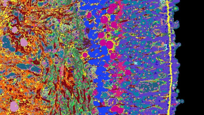
At the heart of the methods the scientists used for their approach was the 4i technology: iterative indirect immunofluorescence imaging. This new imaging technique can visualize several dozen proteins in a thin tissue section at high resolution using fluorescence microscopy. The 4i technology was developed a few years ago by Lucas Pelkmans, a professor at the University of Zurich and coauthor of the study that has just been published in the scientific journal Nature Biotechnology. It is in this study that the researchers applied this method to organoids for the first time.
Typically, researchers use fluorescence microscopy to highlight three proteins in a tissue, each with a different fluorescent dye. For technical reasons, it is not possible to stain more than five proteins at a time. In 4i technology, three dyes are used, but these are washed from the tissue sample after measurements have been taken, and three new proteins are stained. This step was performed 18 times, by a robot, and the process took a total of 18 days. Lastly, a computer merges the individual images into a single microscopy image on which 53 different proteins are visible. They provide information on the function of the individual cell types that make up the retina; for example, rods, cones, and ganglion cells.
The researchers have supplemented this visual information of retinal proteins with information on which genes are read in the individual cells.
High Spatial and Temporal Resolution
The scientists performed all these analyses on organoids that were of different ages and thus at different stages of development. In this way, they were able to create a time series of images and genetic information that describes the entire 39-week development of retinal organoids. “We can use this time series to show how the organoid tissue slowly builds up, where which cell types proliferate and when, and where the synapses are located. The processes are comparable to those of retinal formation during embryonic development,” says Gray Camp, a professor at the University of Basel and a senior author of this study.
The researchers published their image information and more findings on retinal development on a publicly accessible website: EyeSee4is.
Further Tissue Types Planned
So far, the scientists have been studying how a healthy retina develops, but in the future, they hope to deliberately disrupt development in retinal organoids with drugs or genetic modifications. “This will give us new insights into diseases such as retinitis pigmentosa, a hereditary condition that causes the retina’s light-sensitive receptors to gradually degenerate and ultimately leads to blindness,” Camp says. The researchers want to find out when this process begins and how it can be stopped.
Treutlein and her colleagues are also working on applying the new detailed mapping approach to other tissue types, such as different sections of the human brain and to various tumor tissues. Step by step, this will create an atlas that provides information on the development of human organoids and tissues.
Reference: “Multimodal spatiotemporal phenotyping of human retinal organoid development” by Philipp Wahle, Giovanna Brancati, Christoph Harmel, Zhisong He, Gabriele Gut, Jacobo Sarabia del Castillo, Aline Xavier da Silveira dos Santos, Qianhui Yu, Pascal Noser, Jonas Simon Fleck, Bruno Gjeta, Dinko Pavlinić, Simone Picelli, Max Hess, Gregor W. Schmidt, Tom T. A. Lummen, Yanyan Hou, Patricia Galliker, David Goldblum, Marton Balogh, Cameron S. Cowan, Hendrik P. N. Scholl, Botond Roska, Magdalena Renner, Lucas Pelkmans, Barbara Treutlein and J. Gray Camp, 8 May 2023, Nature Biotechnology.
DOI: 10.1038/s41587-023-01747-2
Never miss a breakthrough: Join the SciTechDaily newsletter.
Follow us on Google and Google News.