Researchers have developed a new platform that can replicate different types of blood vessel structures.
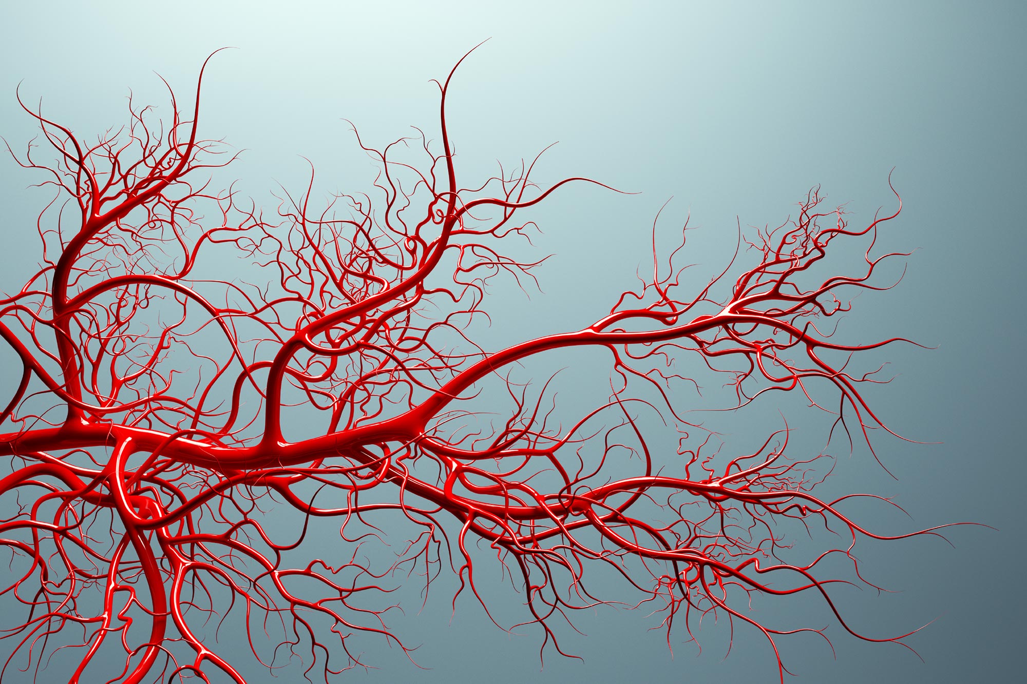
Blood vessels are less like straight pipes and more like a crowded city road map, with turns, forks, and sudden choke points that can change how traffic moves. For a long time, many lab built vessel models skipped that complexity and relied on simple, straight channels, even though real vessels rarely behave that neatly.
Researchers in the Department of Biomedical Engineering at Texas A&M University are trying to close that gap with a customizable vessel-chip method. The goal is to recreate the kinds of shapes that matter in disease, so experiments on blood flow and potential treatments reflect what happens in the body more closely and can better support drug discovery.
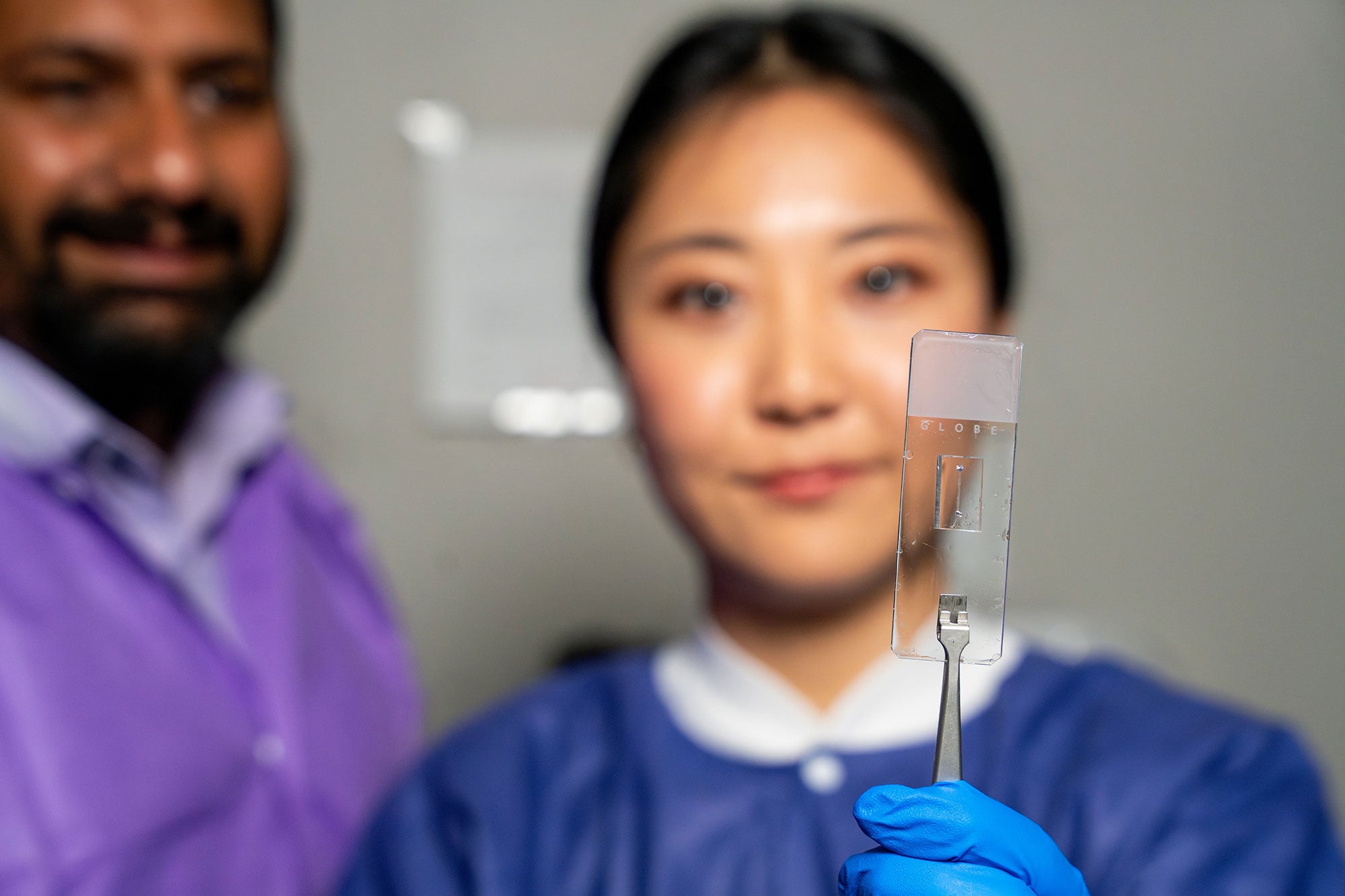
Vessel-chips are engineered microfluidic devices that mimic human vasculature on a microscopic scale. Instead of studying blood flow in animals or oversimplified lab setups, scientists can use these chips to examine how fluid forces move through vessel-like structures in a controlled environment. Because the design can be tailored, the platform can also support patient-focused studies, which is especially useful when small differences in anatomy may affect how disease develops or how a therapy performs.
Jennifer Lee, a biomedical engineering master’s student, joined Dr. Abhishek Jain’s lab and designed an advanced vessel-chip that could reproduce real variations in vascular structure. That matters because vessel geometry can reshape the physical forces acting on the vessel wall. Those forces, including shear stress, influence how cells lining blood vessels respond, and they can contribute to the conditions in which damage begins or disease progresses.
“There are branched vessels, or aneurysms that have sudden expansion, and then stenosis that restricts the vessel. All these different types of vessels cause the blood flow pattern to be significantly changed, and the inside of the blood vessel is affected by the level of shear stress caused by these flow patterns,” Lee said. “That’s what we wanted to model.”
Building on Earlier Innovations
Lee’s research was published in Lab on a Chip and followed earlier work from her mentor, Dr. Tanmay Mathur, a former biomedical engineering graduate student who previously created a straight vessel-chip. Lee and Mathur conducted their studies in the Bioinspired Translational Microsystems Laboratory under Jain, an associate professor and Barbara and Ralph Cox ’53 faculty fellow in the biomedical engineering department. The new research will also appear on the cover of the May 2025 issue of the journal.
“We can now start learning about vascular disease in ways we’ve never been able to before,” Jain said. “Not only can you make these structures complex, you can put actual cellular and tissue material inside them and make them living. These are the sites where vascular diseases tend to develop, so understanding them is critical.”
Lee entered Jain’s lab as an undergraduate honors student seeking research experience. Lee said she didn’t know much about the organs-on-a-chip platform but became interested in its potential to shape the future of medicine. As she transitioned into graduate studies, Lee developed an interest in vessel-chips and joined the Master of Science fast-track program to continue her newfound passion for research.
“Jennifer demonstrated perseverance, curiosity, and creativity, and started taking up research projects very quickly. Our fast-track program enables students like Jennifer to take on sort of high-impact, high-risk research and not just do a science project, but take it all the way to its outcome and get it published,” Jain said.
Expanding the Living Vessel Model
While this iteration of the vessel-chip improves physiological relevance, Jain and Lee hope to expand their research by including various cell types. Lee’s research currently only uses endothelial cells — or cells that make up the lining of the blood vessel — but they hope to include other cells to see the effects of their interactions with each other and blood flow.
“We are progressing and creating what we call the fourth dimensionality of organs-on-a-chip, where we not only focus on the cells and the flow, but this interaction of cells and flow in more complex architectural states, which is a new direction in the field,” Jain said.
In addition to research experience, Lee has gained a multitude of soft skills and the ability to apply concepts learned in class to a real-world experience.
“It’s such a good environment to interact with not only peers but also graduate students and postdoctoral researchers,” she said. “You’re able to learn teamwork and communication, work ethic, and just trying different things out. I think it’s such a valuable experience that students have available. We have such good faculty research labs.”
Reference: “Vascular architecture-on-chip: engineering complex blood vessels for reproducing physiological and heterogeneous hemodynamics and endothelial function” by Jennifer D. Lee, Ankit Kumar, Tanmay Mathur and Abhishek Jain, 5 March 2025, Lab on a Chip.
DOI: 10.1039/D4LC00968A
The research was supported by the U.S. Army Medical Research Program, NASA, Biomedical Advanced Research and Development Authority, National Institutes of Health, U.S. Food and Drug Administration, National Science Foundation, and Texas A&M University Office of Innovation Translational Investment Funds.
Never miss a breakthrough: Join the SciTechDaily newsletter.
Follow us on Google and Google News.
2 Comments
Great progress.
What I find mysterious, is how angiogenesis manages to direct arteries and veins to meet up at capillaries to complete the circulation loop.
Doctors need to return to “SEA SALT WATER” for TRANSFUSIONS
.
Stop blood transfusions